Presentation
Multiple scalp swellings.
Patient Data
Age: 10 months
Gender: Female
From the case:
Eosinophilic granuloma of the skull


Download
Info

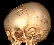
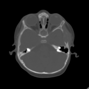
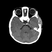
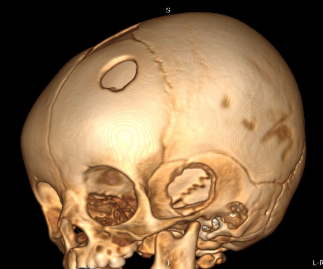
Two osteolytic lesions are seen in the left temporal and left frontal bones. They have bevelled edges with wider bone destruction in the outer than the inner skull table.
Case Discussion
Eosinophilic granuloma or histiocytosis X is most often seen in children aged 1 to 15 with the peak incidence from 1 to 4 years of age.
Characteristic appearance in the skull:
- punched out lytic lesions without sclerotic rim
- double contour or bevelled edge appearance may be seen due to asymmetrical involvement of the inner versus the outer table (hole within a hole sign), best seen in the VR images.


 Unable to process the form. Check for errors and try again.