
Presentation
Large palpable abdominal mass on physical exam. Refractory hypertension.
Patient Data
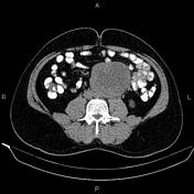

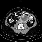
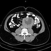

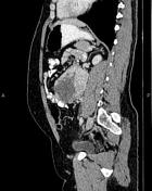
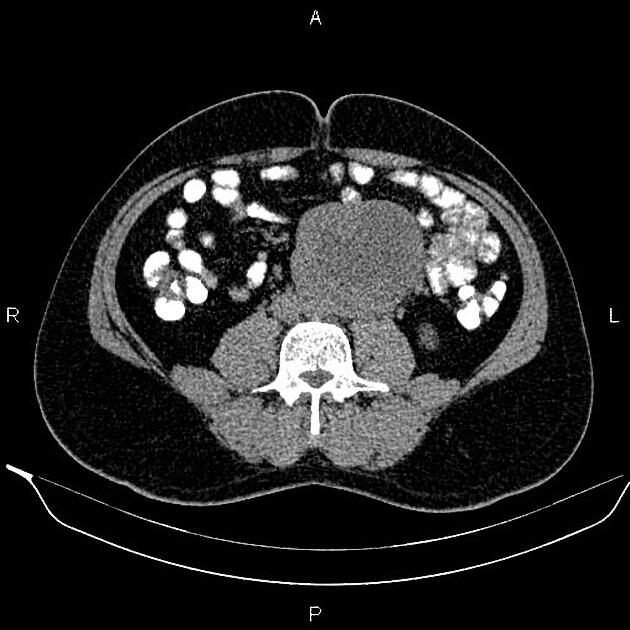
A 110×80×90 mm well-defined hetero enhancing mass is noted at the left para aortic region which shows a large area of internal necrosis.
Case Discussion
Regarding imaging findings, large necrotic lymphadenopathy, extra-gonadal germ cell tumor, extra-adrenal paraganglioma, and retroperitoneal sarcoma could be suggestive in the differential list.
The patient underwent retroperitoneal mass resection.
Histopathology:
A "retroperitoneal mass" consists of a piece of oval tissue measuring 9x8x6 cm; on a cutting cut, the surface is tan-creamy, heterogeneous, with necrotic and hemorrhagic areas.
IHC study result: Positive for Synaotophysin, Chromogranin, and S100, and negative for CKAE1/AE3. Ki67 is positive in 1-2% of tumor cells.
Diagnosis: Extra-adrenal paraganglioma, retroperitoneal mass.
Nuclear pleomorphism: Not identified
Mitotic figures are not easily found.
Dominant pattern: Nest and insular pattern
Necrosis: Present
Lymphovascular invasion: Not identified
Capsular invasion: Not identified
Margins: Free from tumor
Pathologic stage classification (pTNM, AJCC 8th edition): pT2
Paragangliomas are rare neuroendocrine tumors arising from paraganglia and abdominopelvic extra-adrenal paragangliomas are usually originated from organ of Zuckerkandl (as in this case) or bladder base.
It should notice that malignancy is defined as evidence of metastases and histopathologically there are no reliable markers of malignant potential.


 Unable to process the form. Check for errors and try again.