
Presentation
Staging and better characterization of a retroperitoneal mass detected on abdominal ultrasonography.
Patient Data
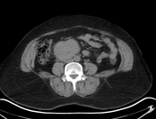

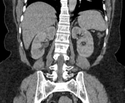
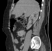
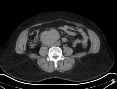
A well-defined heterogeneously enhancing retroperitoneal soft tissue mass with areas of cystic necrosis inside is seen along the rightward aspect of the abdominal aorta (opposite L3 & L4 vertebral bodies) with a small portion draping along its anterior aspect near the level of origin of the IMA. The mass is also seen compressing the anterior aspect of the infra-hepatic IVC, contacting the right psoas muscle posteriorly as well as the 3rd part of the duodenum superiorly. It measures about 5.8 x 7 x 6 cm.
Case Discussion
This mass was biopsied and proved to be extra-adrenal pheochromocytoma (paraganglioma) on histopathological examination.
On CT, the mass appeared to be confined to the retroperitoneum with vascular contact to the aorta and IVC with compression of the latter.


 Unable to process the form. Check for errors and try again.