
Presentation
Abdominal pain, distension, and vomiting - two days
Patient Data

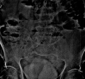
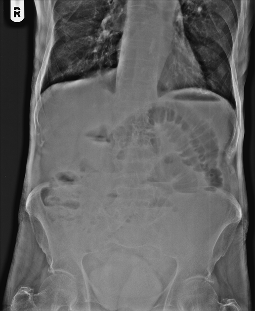
Multiple small bowel air-fluid levels. No free gas under the domes of the diaphragm. The magnified cropped image shows an obliquely oriented short string of small lucencies over the sacrum, a string of pearl sign.
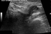

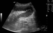
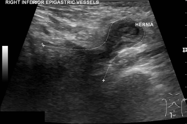
Right groin irreducible hernia containing small bowel. A narrow neck of the hernia (about 7 mm). Long-axis images show a caudally directed hernia passing deep to the right inguinal canal. The hernia neck is a little away from the inferior epigastric vessels. The short-axis images below the inguinal canal show the hernia sac lying medial to the collapsed femoral vein. Herniated small bowel wall edema with preserved vascularity. Right inguinal hernioplasty status. No hernia extension in the right inguinal canal.
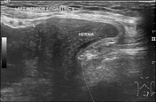
Left groin reducible hernia containing small bowel with wide hernia neck. It passes deep/ medial to the inferior epigastric vessels in the left inguinal canal. The short-axis images show a wide gap between the hernia sac and the non-collapsed femoral vein.
Multiple dilated small bowel loops with hyperperistalsis
No ascites.
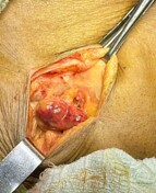
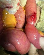

The first photo shows the right femoral hernia containing a trapped bowel loop. The second photo shows an affected small bowel segment after hernia reduction. There are constriction marks and wall hemorrhage. It was viable. No bowel resection was required.
Case Discussion
The case shows a right femoral hernia and a left inguinal hernia. Small bowel obstruction due to narrow neck of the right femoral hernia which was surgically treated within a few hours after the ultrasound.
Intraoperative photos courtesy: operating surgeon Dr. Sanjay Prasad.


 Unable to process the form. Check for errors and try again.