
Presentation
Slowly growing painless swelling over the volar aspect of the the right wrist and palm.
Patient Data
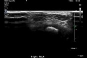


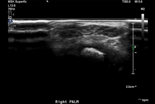
The right median nerve is swollen and enlarged at the level of the wrist and right palm. The nerve fascicles are enlarged and hypoechoic surrounded by echogenic fat tissue giving cable-like appearance on transverse images. No abnormal vascularity of the enlarged nerve.
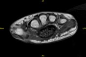
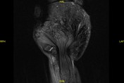

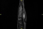
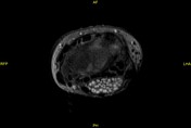

The median nerve is swollen with cable-like appearance on axial images and spaghetti-like appearance on the coronal images. The nerve fascicles are of intermediate signal on T1, high signal on T2 and show post-contrast enhancement. The nerve fascicles are interspersed with connective tissue showing signal drop on fat suppression images.
Case Discussion
Fibrolipomatous hamartomas are benign tumors of the nerves. The median nerve is the most commonly involved. The appearance of cable-like and spaghetti-like enlargement of the nerve that are easily appreciated on MRI, are considered pathognomonic allowing for non-invasive diagnosis of this pathology.


 Unable to process the form. Check for errors and try again.