
Presentation
Right side eye proptosis and hypotropia and right side facial deformity and upper eyelid bulge.
Patient Data
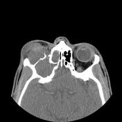

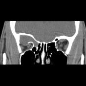
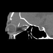

The large expansile relatively hypodense mass-like lesion within the right frontoethmoidal sinus extended within the right upper orbital cavity extraconal through a large bone defect is seen that has a remarkable impression on related extraocular muscle and eye globe and has led to pronounced proptosis and hypotropia. Fluid-filled expansion of all parts of the frontal sinus is also seen. Focal encephalomalacia in the left side posterior frontal lobe and adjacent dilated frontal horn are also noted.
Case Discussion
The case illustrates a large fronto-ethmoidal mucocele extended within the right orbital cavity and caused pronounced proptosis and hypotropia. The frontal sinus is the most common location of mucoceles of paranasal sinuses and one of the major complications of frontal sinus mucocele is orbital cavity invasion. The diagnosis of this case was based on MDM discussion, typical imaging features, and surgical findings.


 Unable to process the form. Check for errors and try again.