
Presentation
Melena.
Patient Data
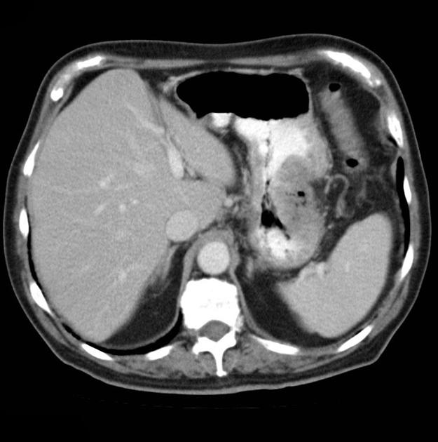
CT of the abdomen demonstrates a mass arising from the greater curvature of the body of the stomach. It is centrally ulcerated with locules of gas and contrast seen within it, but without evidence of perforation. It is a focal abnormality with thick heaped-up shoulders.
At the splenic hilum there appears to be an enlarged lymph node (this should be confirmed on thin slice and coronal reformats). No convincing evidence of metastatic disease.
The left kidney is atrophic and contains a number of what appear to be hyperdense cysts (which should be confirmed on ultrasound). The right kidney has a large simple cyst.
This patient went on to have a total gastrectomy after the stomach tumor was identified on gastroscopy.
HISTOLOGY
MACROSCOPIC DESCRIPTION: The specimen is a total gastrectomy. Arising from the body of the mucosa at the anterolateral aspect is a large friable tan centrally ulcerated round polypoid tumor mass 65 mm in diameter with markedly heaped up edges. At the site of the tumor the serosal surface shows marked indrawing, roughening and there is adherent fat. Macroscopically the tumor appears to extend through the wall to the serosal surface.
MICROSCOPIC DESCRIPTION: The tumor is adenocarcinoma with abundant gland formation and mucin secretion (so-called intestinal type). It infiltrates through the full thickness of stomach wall into adjacent fat. The surface is ulcerated and covered by abundant necrotic and inflammatory debris with abundant bacteria. Focally the tumor cells infiltrate as large solitary atypical cells and although the bulk of the tumor is moderately differentiated, at worst the tumor is considered poorly differentiated.
DIAGNOSIS: moderately and focally poorly differentiated adenocarcinoma
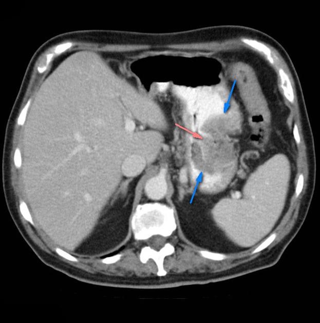
Heaped-up margins of the mass (blue arrows) with central ulceration (pink arrow).
Case Discussion
This tumor demonstrates typical features of a large stomach adenocarcinoma, with heaped up edges and central ulceration, which is a feature of up to 70% of gastric adenocarcinomas.


 Unable to process the form. Check for errors and try again.