
Presentation
This patient came to the hospital emergency room with complaints of pain and abdominal distension without nausea or vomiting.
Patient Data


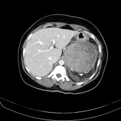
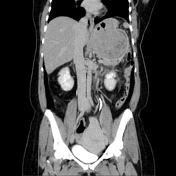
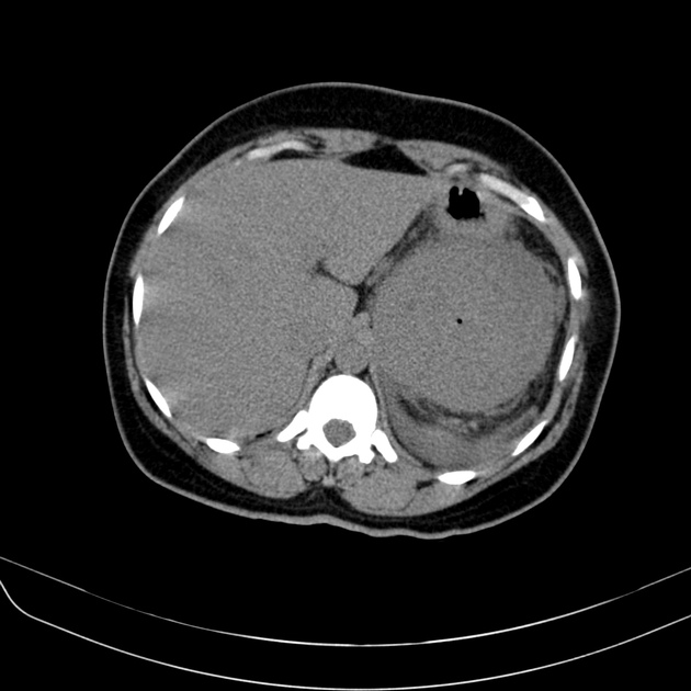
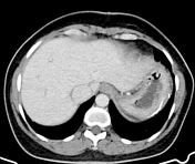
Contrast-enhanced computed tomography (CECT) revealed a heterogeneous solid expansive mass with irregular enhancement located in the upper left quadrant of the abdomen, adjacent to the gastric greater curvature. There were no cleavage planes between the tumor and the stomach, leading to extrinsic compression of the gastric chamber, displacing it in a cephalic direction. This tumor also exerted pressure on the splenic angle of the colon, although there are cleavage planes between these structures. The dimensions of the mass were approximately 10 cm anteroposteriorly x 9.5 cm transversely x 8.0 cm longitudinally.
Additionally, the imaging demonstrated features consistent with calculous cholecystopathy and involvement of uterine leiomyomatosis.
Impression: The primary diagnostic consideration is a solid expansive gastric lesion with exophytic growth, which is most suspicious for a gastric gastrointestinal stromal tumor (GIST).
The patient went on to have a surgical procedure with vertical partial gastrectomy and en bloc splenectomy, with preservation of the pancreas, duodenum, and colon. In addition, she had cholecystectomy for cholelithiasis and a resection of the giant uterine leiomyoma (myomectomy).
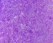
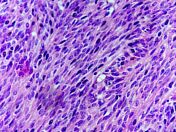


Histology report:
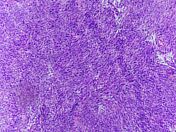
The sample contains mature adipose tissue and tumor fragments represented by atypical spindle cell neoplasm, histologically compatible with gastrointestinal stromal tumor (GIST), low-grade spindle cell subtype - 5 mitoses / 50 CGA.
Conclusion: Fusocellular neoplasia - GIST tumor.


Follow-up CECT approximately thirty months after surgery demonstrated post-surgical changes along with the greater curvature of the gastric body, with resection of the mass and splenectomy. A hypodense fluid layer is present in the splenic bed, which persists unchanged in subsequent CT scans, likely of residual post-surgical nature.
Surgical absence of the gallbladder.
The uterus has enlarged dimensions and signs of leiomyomatous involvement.
Impression: The control tomographic study demonstrates post-surgical changes along the greater curvature of the gastric body, with no signs of recurrent lesions in this region.
Case Discussion
Gastrointestinal stromal tumors (GISTs) are neoplasms originating in the digestive tract, typically identifiable through computed tomography imaging 1-4. This case illustrates the characteristic radiological features associated with GISTs located in the stomach.
Case courtesy
Felipe Coelho de Aguiar, MD - oncologist
Raimundo Neto, MD – surgeon
Horácio Fittipaldi, MD - pathologist
Antonio Rodrigues de Aguiar Neto, MD - radiologist


 Unable to process the form. Check for errors and try again.