
Presentation
Palpable abdominal mass on physical exam.
Patient Data
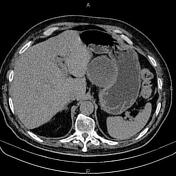

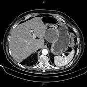

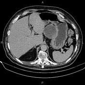

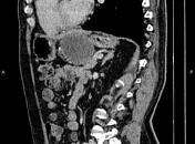

A 72×65 mm mass with internal cystic/ necrotic changes is seen between the left liver lobe and gastric lesser curvature originating from the stomach wall.
A few non-enhanced simple cortical cysts are seen in both kidneys. Two small stones less than 4 mm are seen in the left kidney.
A 15 mm nodule with a mean attenuation value of 8 HU and rapid washout on post contrast images is noted at the left adrenal gland inferring benign adenoma.
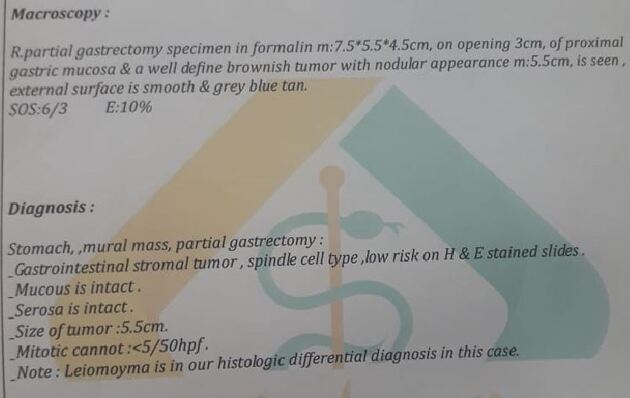
The patient underwent partial gastrectomy and histopathology evaluation confirmed gastrointestinal stromal tumor (GIST).
Case Discussion
It is difficult to differentiate a benign from a malignant GIST on imaging, but exogastric growth, diameter >5 cm, central necrosis, and extension to other organs suggest malignant transformation.


 Unable to process the form. Check for errors and try again.