
Presentation
Sever pain in left ankle with no history of trauma.
Patient Data
Age: 20 years
Gender: Male
From the case:
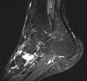
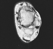
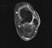
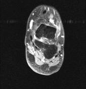
Giant cell tumor with CT guidance biopsy

Download
Info
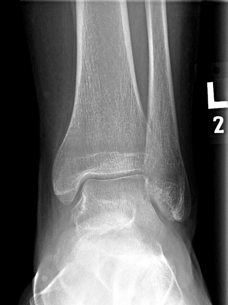
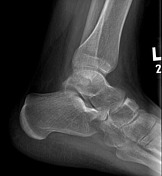
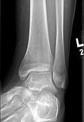
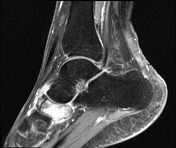
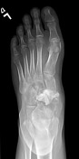
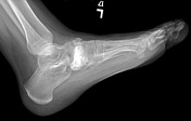
An expansile subtle lytic lesion in the navicular bone.
From the case:
Giant cell tumor with CT guidance biopsy

Download
Info

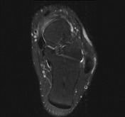
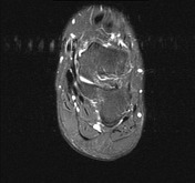
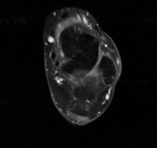
An expansile lesion in the navicular bone with internal and superior soft tissue components.

Download
Info
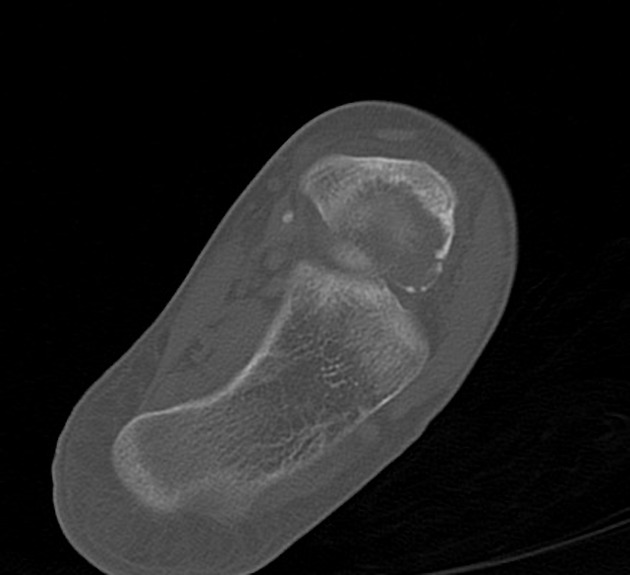
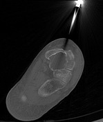
Bone window CT scan images show the epicenter of the mass within the navicular bone.
From the case:
Giant cell tumor with CT guidance biopsy
Download
Info

Status post packing of the navicular bone.


 Unable to process the form. Check for errors and try again.