
Presentation
Expressive and receptive dysphasia and alexia
Patient Data
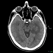

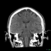
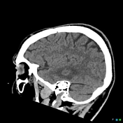
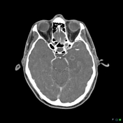
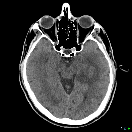
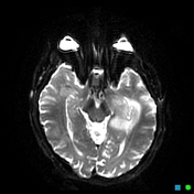
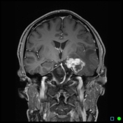
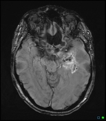
Left temporal lobe vasogenic edema, particularly in the parahippocampal gyrus and mesial temporal lobe. There are two discrete ring-enhancing lesions. The first is an intra-axial ring enhancing mass lesions up to 11mm adjacent to the anterior left hippocampus. The second lesion measures up to 14mm anteroposterior measurement sits on the lateral margin of the left hippocampus. There is local mass effect with sulcal effacement in the mesial left temporal lobe. Otherwise no other intracranial abnormality is found. No intracranial hemorrhage. No parenchymal changes to suggest infarction. No bony abnormalities.

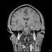
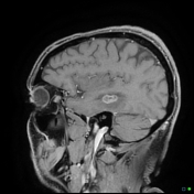
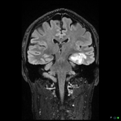
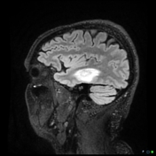
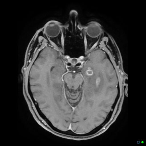
There are three ring-enhancing lesions in the left medial temporal lobe measuring 20 x 9mm, 10 x 11mm and 3 x 4mm respectively. They are located with in the hippocampal region of the temporal lobe and abut the temporal horn of the lateral ventricle. The lesions are suspicious for a malignant process, metastatic malignancy or multifocal glioblastoma. No lesions elsewhere identified on these images. No intracranial hemorrhage.

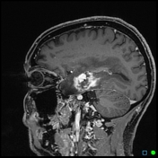
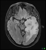
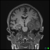
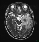
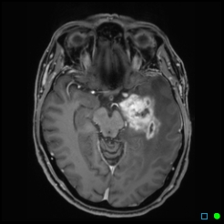
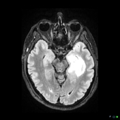
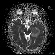
Two irregular heterogeneously enhancing hemorrhagic lesions within the medial left temporal lobe. Both demonstrate increase in size with the largest lesion now measuring 36mm anteroposteriorly. Interval progression in FLAIR signal abnormalities which is involving essentially the whole left temporal lobe and extending into the left frontal parietal and occipital lobes. Associated effacement of the left lateral ventricle is present which is new since the previous study. 4mm midline shift to the right. The above findings are consistent with disease progression.
Case Discussion
Diagnosis of left mesial temporal lobe WHO grade 4 glioblastoma (IDH wild type) on biopsy (histology below) without resection. Treatment with definitive chemoradiotherapy of 60Gy and temozolomide. High dose dexamethasone use for vasogenic edema related symptoms. Disease progression despite ongoing adjuvant therapy with temozolomide.
Histology Report:
Left temporal lobe biopsy:
The sections show fragmented pieces of moderately cellular tumor with moderate nuclear atypia, frequent mitotic figures and areas of necrosis.
Immunohistochemical stains have been performed with the following results:
IDH1-R132H: negative
P53: wild type
ATRX: retained
The immunohistochemical features are suggestive of a primary glioblastoma.
Summary: Glioblastoma, IDH negative by immunohistochemistry, WHO grade 4.


 Unable to process the form. Check for errors and try again.