
Presentation
Visual disturbances, headaches and expressive dysphasia. Visual disturbances thought to be secondary to lymphoid hyperplasia within the right the right orbit. Past medical issues include: seizures, HBV, HCV, crack cocaine and speed use, pack-a-day smoker.
Patient Data

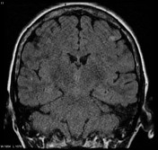
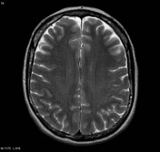
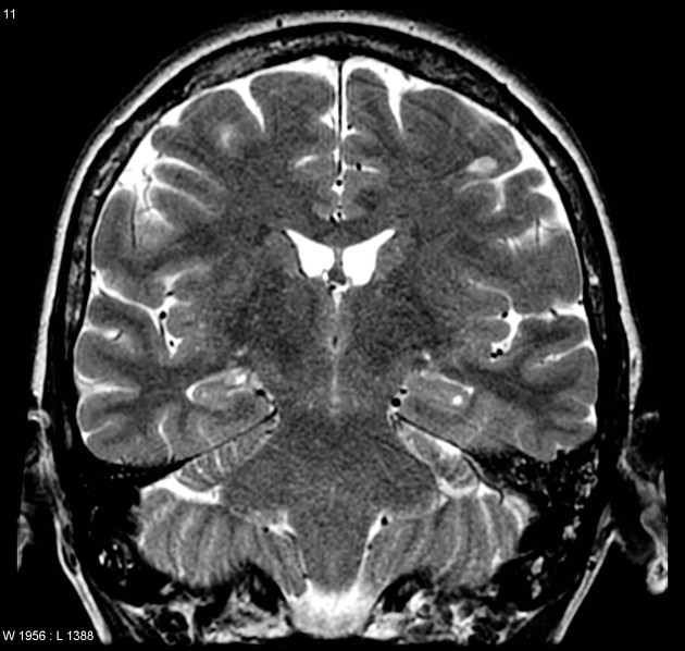
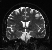
A small subcortical region of high T2 signal is seen in the left frontal lobe.
The patient was lost to follow-up for 5 years and represented with a further seizure. A CT demonstrated an abnormality which prompted further MRI imaging.

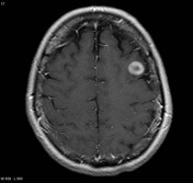
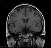
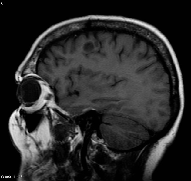
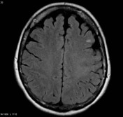
The region has increased in size and now vividly enhances.
The patient refused treatment and again failed to attend follow-up visits. They were homeless and unable to be located. 6 months later they presented with increasing headaches and seizures.

A peripherally enhancing lesion is located within the left frontal lobe.
Case Discussion
Finally, the patient consented to surgery. Histology demonstrated a glioblastoma (GBM).
It is uncommon to watch a brain tumor grow for 5 years, but when complicated psychosocial issues are present following up patients or convincing them to have treatment can be difficult.
Note: This case predates the recent (2016) revision WHO classification of CNS tumors and IDH status is not available. Although with such a protracted documented history this is likely a secondary GBM and thus likely IDH mutated, as this has not been established this tumor would now be classified as a glioblastoma NOS.


 Unable to process the form. Check for errors and try again.