
Presentation
Headache, decreased vision and lack of concentration of 3 weeks duration.
Patient Data
Age: 55 years
Gender: Male
From the case:
Glioblastoma NOS


Download
Info
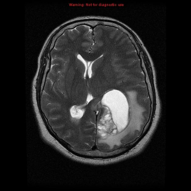
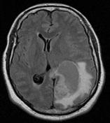
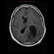
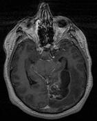
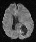
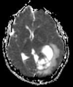
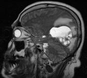
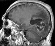
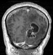
MRI brain demonstrates a left occipital lobe intra-axial mixed solid and cystic lesion associated with vasogenic edema and mass effect.
Case Discussion
The patient went on to have surgery, and histology confirmed the diagnosis of glioblastoma.
Note: IDH mutation status is not provided in this case and according to the current (2016) WHO classification of CNS tumors, this tumor would, therefore, be designated as a glioblastoma NOS.


 Unable to process the form. Check for errors and try again.