
Presentation
Aphasia with gait disturbance
Patient Data
Age: 60 years
Gender: Female
From the case:
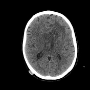
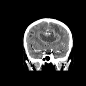
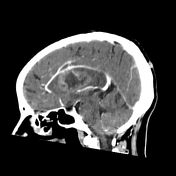
Glioblastoma NOS - butterfly appearance


Download
Info

There is a large midline lobulated mass crossing the corpus callosum into the white matter of the frontal lobes with "butterfly" appearance. It shows thick and irregular margins, isodense to the gray matter on non-contrast images with irregular and heterogeneous enhancement on postcontrast images. The center of this mass appears irregular, hypodense (central necrosis). Surrounding vasogenic edema is noted as well as a mass effect on the frontal horns.
Multiple subcutaneous sebaceous cysts are noted.
Case Discussion
CT features highly suggestive of a high-grade butterfly glioma (glioblastoma NOS)


 Unable to process the form. Check for errors and try again.