
Presentation
This elderly patient presenting with short term memory loss, confusion and a change in personality.
Patient Data


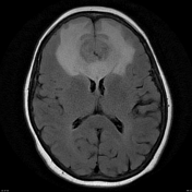
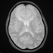
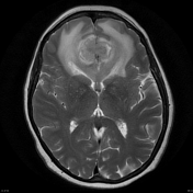
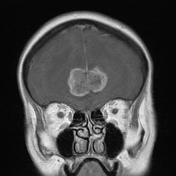
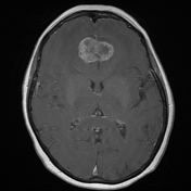
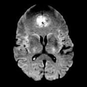
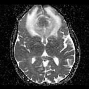
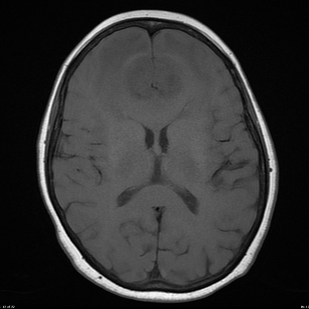
In the anterior midline is a rounded mass with heterogeneous contrast enhancement, which is more prominently peripheral. There is very extensive vasogenic edema, also involving the genu of the corpus callosum, which also appears enlarged.
Case Discussion
This is a difficult case, due to the location of the tumor.
The finding which draws one away from the diagnosis of a GBM is the midline location and the fact that the anterior cerebral arteries are located posterior to the mass and anterior to the corpus callosum. Thus this is not a genu mass, and one could be mistaken in thinking that it arose from the falx. One should remember that the falx, this far anteriorly, is usually a thin strip descending towards the crista galli.
The patient was taken to theater and resection performed. The mass was more reminiscent of a glioblastoma (GBM) and frozen section confirmed the diagnosis (subsequently further confirmed with histology).
Histology
The sections show features of a densely cellular astrocytic tumor. The tumor cells show elongated, angulated and hyperchromatic nuclei. Some appear pleomorphic xanthoastrocytoma-like, being more epithelioid with abundant amounts of cytoplasm. Scattered mitotic figures are identified. There are foci of endothelial cell hyperplasia. Areas of palisaded necrosis are present. The features are those of glioblastoma multiforme. The tumor cells are GFAP and nestin positive. The Ki-67 index is about 30%.
FINAL DIAGNOSIS: Glioblastoma (WHO Grade IV).
Note: IDH mutation status is not provided in this case and according to the current (2016) WHO classification of CNS tumors, this tumor would, therefore, be designated as a glioblastoma NOS.


 Unable to process the form. Check for errors and try again.