
Presentation
Right side proptosis and orbital pain for five months.
Patient Data
Age: 40 years
Gender: Female
From the case:
Granular cell tumor

Download
Info

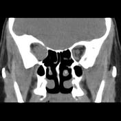
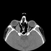
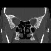
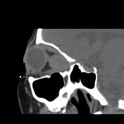
Mass like enlargement of the right orbital medial rectus muscle with axial width up to 22 x 18 mm and right eye proptosis and exodeviation are seen. The impression and deformity on the related orbital posterior medial wall due to the mass lesion are also noted. Pronounced impression on adjacent optic nerve sheath complex is noted.
Case Discussion
The case illustrates the non-contrast MDCT features of pathology-proved benign granular cell tumor or myoblastoma of the medial rectus muscle. Extraocular muscles are a rare location of the tumor and the main treatment is complete surgical excision of the tumor that is not always feasible 1.


 Unable to process the form. Check for errors and try again.