
Presentation
Multiple hepatic focal lesions discovered on sonography.
Patient Data
Age: 2 months
From the case:
Hepatic hemangioendothelioma


Download
Info

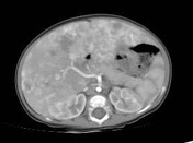
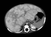
Multiple bi-lobar variable sized hepatic focal lesions are seen showing intense peripheral nodular enhancement on the arterial phase with gradual filling in on the portal-venous and delayed phases.
Reduction of the caliber of the abdominal aorta below the level of celiac branch with celiac trunk and hepatic artery hypertrophy.
Case Discussion
A neonate with multiple hepatic focal lesions showing typical peripheral enhancement and gradual filling in plus reduction in the aortic caliber below the level of celiac branch are diagnostic of hepatic hemangioendothelioma.


 Unable to process the form. Check for errors and try again.