
Presentation
Dyspnea and weight loss.
Patient Data

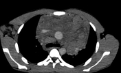

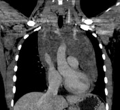
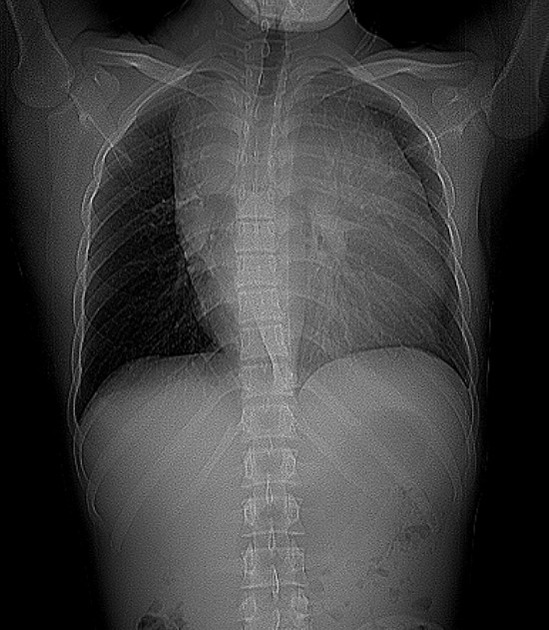
Huge mediastinal soft tissue lesion measuring 12 x 16 x 15.8 cm in AP, SS and CC dimensions respectively, encasing the pericardium and major vessels. The epicardial fat is intact. It encases the ascending aorta, aortic arch, and its main branches as well as the proximal part of the descending aorta. It also encases the pulmonary artery and its main branches. The superior vena cava and brachiocephalic veins are displaced and narrowed. It shows a heterogenous texture comprising nodular soft tissue densities with intervening fat. It shows heterogeneous post-contrast enhancement.
Multiple confluent right paratracheal nodes, left cardiophrenic, right axillary and cervical lymph nodes.
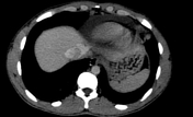
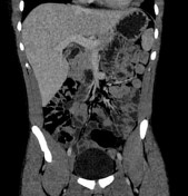

Normal abdominal CT scan. No abnormal abdominal lymph nodes.
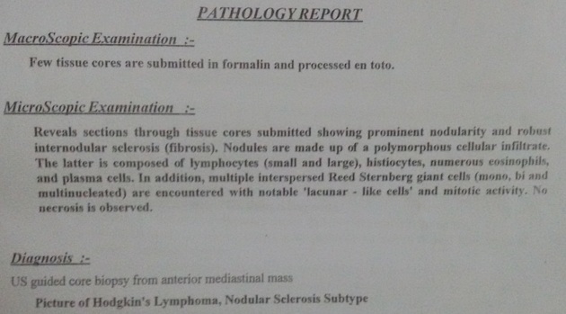
Pathology from mediastinal mass revealed nodular sclerosing subtype of Hodgkin lymphoma
Case Discussion
This case of pathologically proven nodular sclerosing Hodgkin lymphomam presented as a huge mediastinal soft tissue mass lesion with mediastinal lymph nodes as well as cervical and right axillary lymph nodes. No abdominal involvement.
Nodular sclerosing Hodgkin lymphoma is a type of primary mediastinal lymphoma. Approximately 60% of all Hodgkin lymphoma involves the mediastinum at presentation.


 Unable to process the form. Check for errors and try again.