
Presentation
Right hip and low back pain.
Patient Data
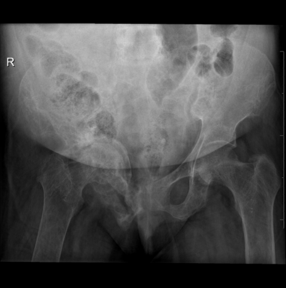
Plain x-ray AP view of the pelvis shows:
osteopenic texture of the radiographed bones
marked subperiosteal bone erosions with slight expansile osteolysis of the right hip bone more severe at the superior and inferior pubic rami
subsequent right protrusio acetabuli


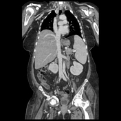
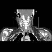
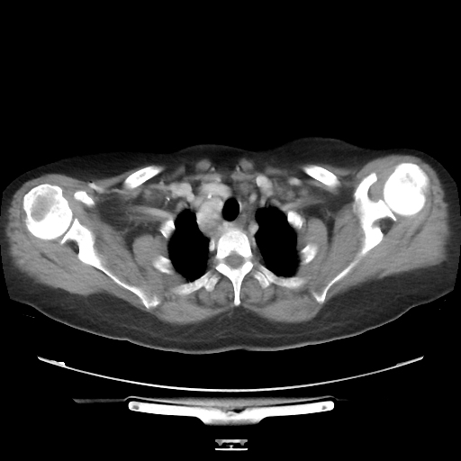
There is a well-defined heterogeneously enhanced soft tissue mass lesion posteroinferior to the right thyroid lobe with retrosternal extension. No calcification seen. Appearances suggest a right parathyroid adenoma.
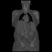
There is diffuse osteopenia involving the visualized skeleton. Evidence of advanced bone resorption, subperiosteal erosions as well as bony erosions are noted, more advanced at the pelvic bones including the ilium, acetabulum, sacral and hip bones. Right hip acetabulum protrusion is noted with an associated joint effusion.
The spine shows diffuse osteopenia with reduced vertebral height and bony erosions at L2.
The overall CT picture is impressive primary hyperparathyroidism due to a right inferior parathyroid adenoma.
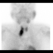

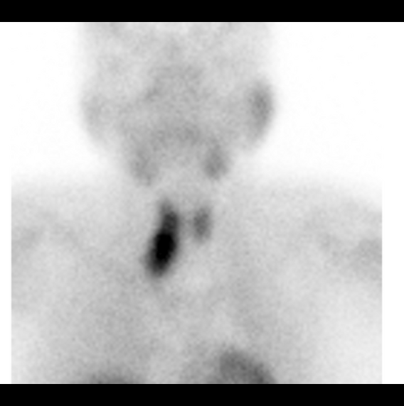
99mTc-Sestamibi nuclear scan shows intense radiotracer uptake at the right lower neck.
Coronal SPECT/CT confirms the location of the parathyroid adenoma related to the mass previously detected by contrast-enhanced CT.
Case Discussion
Laboratory investigation supported the diagnosis at the time of CT scan. Elevated serum Ca++ and elevated levels of parathormone (PTH) were observed.
Surgical management by selective right inferior parathyroidectomy was performed. Histopathology confirmed the diagnosis of parathyroid adenoma.


 Unable to process the form. Check for errors and try again.