
Presentation
Of Asian origin, abdominal pain
Patient Data
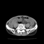

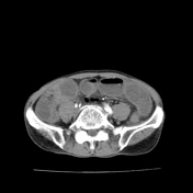
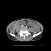

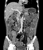
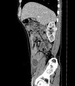

CT findings show dilated loops of small bowel and gas-fluid levels, consistent with small bowel obstruction.
The transition point at the terminal ileum is due to circumferential thickening of the wall and the cecum appears shrunken.
Some enlarged mesenteric lymph nodes are seen in the right iliac fossa.
The enterectomy was done to remove the obstruction.
The samples of resected bowel and lymph nodes confirmed typical histopathological findings of tuberculosis: central caseous necrosis surrounded by epithelioid cells and Langhans giant cells, the outermost are lymphocytes.
Case Discussion
The ileocecal region is the commonest site of tuberculosis in the gastrointestinal tract. However, the image findings are not pathognomonic of tuberculosis. The differential diagnosis includes Crohn disease and carcinoma of the cecum. The histopathology confirmed the diagnosis of tuberculosis.


 Unable to process the form. Check for errors and try again.