Presentation
Palpable bulge during the second trimester of pregnancy.
Patient Data


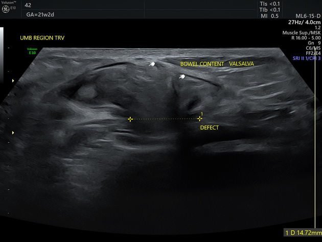
A grayscale sonogram shows a midline defect with a width of 14.72 mm located near the previous laparoscopic scar. Herniation of bowel loops and omental fat on Valsalva can be seen.
The cine clip showed real-time herniation upon straining and completely reduced upon relaxation.
Case Discussion
The patient has a history of laparoscopic appendectomy performed 10 months prior. The swelling has progressively enlarged with the progression of the current pregnancy. The differential diagnosis for an umbilical swelling in pregnancy includes umbilical hernia, rectus diastasis, uterine leiomyoma, and anterior abdominal wall tumors. Ultrasound is the imaging modality of choice due to the patient's gravid status 1.


 Unable to process the form. Check for errors and try again.