
Presentation
Mass in right hypochondrium since birth.
Patient Data


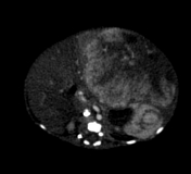
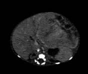
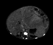
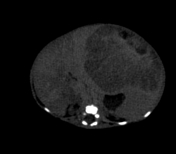
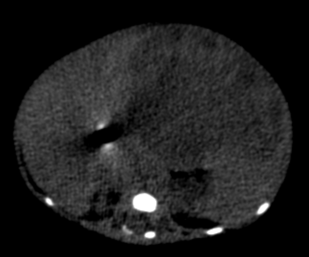
A large mass lesion is seen in the left lobe of liver, which shows peripheral hyperenhancement on arterial phase and gradual filling in on venous and delayed phase.
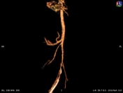
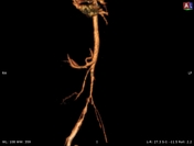
3D MIP images show the characteristic reduction in the aortic diameter below the level of celiac origin, because of the vascular redistribution toward the liver by this large hypervascular mass. This cause celiac trunk and hepatic artery hypertrophy.
Above findings are consistent with Infantile hepatic hemangioma.
Case Discussion
This is a case of histopathological proven infantile hepatic hemangioma, which is a neoplasm of liver caused by the proliferation of endothelial cells. It shows rapid growth in early stages and spontaneous involution.
The term “infantile hepatic hemangioma” is used instead of “hemangioendothelioma” because the clinicobiologic behavior is similar to infantile hemangiomas that occur elsewhere. It differs from adult epithelioid hemangioendothelioma in that it has malignant potential and does not involute.
It produces symptoms such as fetal cardiovascular compromise, hydrops fetalis, hepatomegaly, hemolytic anemia, thrombocytopenia, and unexplained congestive cardiac failure.
Therapeutic options include steroids, embolization, chemotherapy, radiotherapy, surgery, or liver transplantation.


 Unable to process the form. Check for errors and try again.