
Presentation
Abdominal distension.
Patient Data
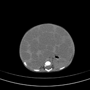

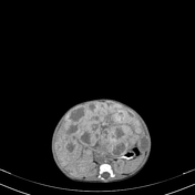
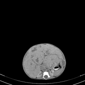

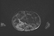
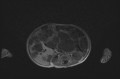
An extensive intra-abdominal mass lesions, which diffusely infiltrate the liver, appearing as multiple hypodense areas in comparison to liver parenchyma. They show peripheral postcontrast enhancement on arterial and gradual filling in on venous phase.
IVC can't be visualized.
Retroperitoneum can't be differentiated from the lesion.
Kidneys and spleen are displaced posteriorly.

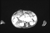
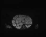
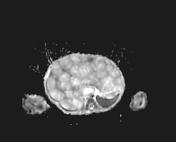
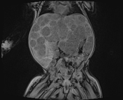
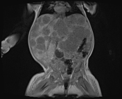
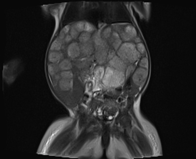
Multiple well-defined spherical masses involving the liver, mostly the left lobe, T1 hypointense and T2 hyperintense relative to the liver, which cause extensive liver enlargement.
Flow voids can be seen in or adjacent to some lesions.
Case Discussion
Histopathology report confirmed infantile hepatic hemangioma.
This case shows near-total replacement of the hepatic parenchyma with many lesions, which caused high-output heart failure due to arteriovenous and portovenous shunting. The patient was treated with propranolol and corticosteroids.


 Unable to process the form. Check for errors and try again.