
Presentation
Lower abdominal pain.
Patient Data
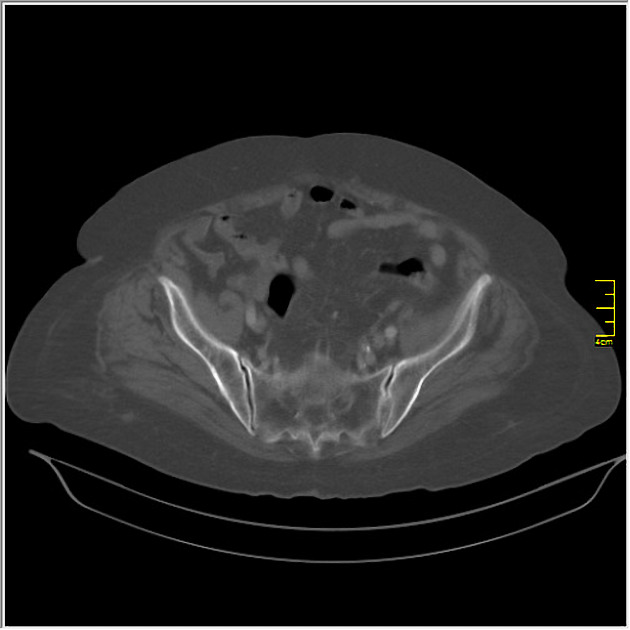
Linear hyperdense foreign body is seen in the sigmoid colon. Notice the colonic wall and adjacent fat tissue inflammation at this region.
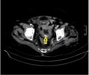
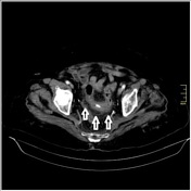
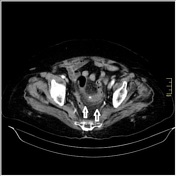
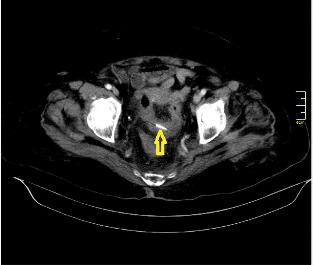
Linear hyperdense foreign body (yellow arrow) is seen in the sigmoid colon. Notice the colonic wall and adjacent fat tissue inflammation at this region (white arrows).
This ingested foreign body (chicken bone) was extracted by colonoscopy.
Note: This case has been tagged as "legacy" as it no longer meets image preparation and/or other case publication guidelines.
Case Discussion
Ingested foreign bodies, such as chicken bones, fish bones and toothpicks, are typically excreted from the digestive tract without any complications. They are infrequently seen in children and elderly people and they may lead to clinical problems such as inflammation, obstruction, perforation or bleeding.


 Unable to process the form. Check for errors and try again.