Presentation
Dizziness and hearing loss.
Patient Data
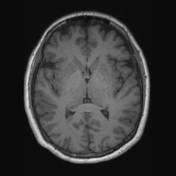

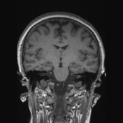

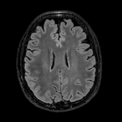

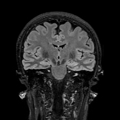

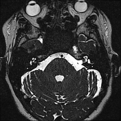

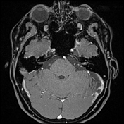

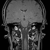

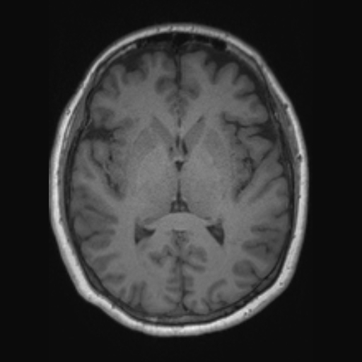
Isointense T1 / T2 signal nodule in the left internal auditory canal with post-contrast enhancement. Broad attachment to the anterior wall of the internal auditory canal.
HISTOPATHOLOGY
MACROSCOPIC:
1. A tiny piece of tissue less than 1 mm.
2. Multiple pieces of pinkish–cream tissue 7 x 5 mm in aggregate.
3. A 1mm tissue fragment. All embedded.
MICROSCOPIC:
The sections show fragments of a fairly paucicellular spindle lesion with collagenous background. Cells are elongated with small, bland nuclei. There is no significant staining for S100. EMA shows patchy staining in some areas. The material is limited however the features are in keeping with a fibroblastic meningioma, WHO grade 1.
DIAGNOSIS:
Internal auditory meatus lesion – features suggestive of a FIBROBLASTIC MENINGIOMA, WHO grade 1.
Case Discussion
Meningiomas very rarely arise purely within the internal auditory canal (IAC). In this region, they more commonly arise at the cerebellopontine angle and extend in the IAC. On imaging, it can be difficult to differentiate from a vestibular schwannoma.


 Unable to process the form. Check for errors and try again.