Presentation
Headache
Patient Data
![](https://prod-images-static.radiopaedia.org/images/64719818/d66e91bebba24e66bffa34037da5175ad42d53c3b3df0d783051bc30b58e6def_thumb.jpeg)
![This study is a stack](/packs/stack-YQKLCKBI.gif)
![](https://prod-images-static.radiopaedia.org/images/64719838/df0206ce5cc22d605329e1b7a02d46e28350b8ea77574681003f98247ec7615d_thumb.jpeg)
![This study is a stack](/packs/stack-YQKLCKBI.gif)
![](https://prod-images-static.radiopaedia.org/images/64719857/2b0fe466c77aaecfb7f1c8bc9e9429a34d6e17b7ceceb3eb3419c5b717a82252_thumb.jpeg)
![This study is a stack](/packs/stack-YQKLCKBI.gif)
![](https://prod-images-static.radiopaedia.org/images/64719878/122d60cff8e6a5397498232e075a162c805166d5d357495cdf02785761a71555_thumb.jpeg)
![This study is a stack](/packs/stack-YQKLCKBI.gif)
![](https://prod-images-static.radiopaedia.org/images/64719892/5977ff016a61e38a4c61d5e3af66c207faa017e8dd3332a56f1eddf657e69270_thumb.jpeg)
![This study is a stack](/packs/stack-YQKLCKBI.gif)
![](https://prod-images-static.radiopaedia.org/images/64719929/189d82528487febf098c0085e64057afaa7a9cf901d20ec1a013ef1e4d3175c7_thumb.jpeg)
![This study is a stack](/packs/stack-YQKLCKBI.gif)
![](https://prod-images-static.radiopaedia.org/images/64720056/d1407e7b0a65594c2187e188d920c614be29a8213fc5d0c16e149f65ad46c99c_thumb.jpeg)
![This study is a stack](/packs/stack-YQKLCKBI.gif)
![](https://prod-images-static.radiopaedia.org/images/64719818/d66e91bebba24e66bffa34037da5175ad42d53c3b3df0d783051bc30b58e6def_big_gallery.jpeg)
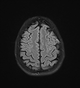
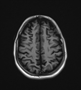
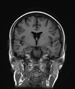
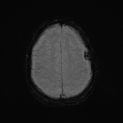
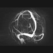
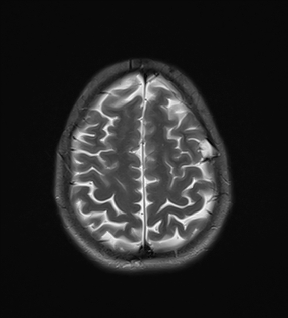
Left parietal calvarial lesion is seen measuring 1.7 cm and displaying low signal on T1, bright signal on T2 and DWIs. Right maxillary sinusitis. Normal MRV.
![](https://prod-images-static.radiopaedia.org/images/64720118/767d732de69314ac7a792aebb53c92b794450d2af0f9f62c6bd3966ea5fb919a_thumb.jpeg)
![This study is a stack](/packs/stack-YQKLCKBI.gif)
![](https://prod-images-static.radiopaedia.org/images/64720133/05fa485c534d12acac1e1c3dc41b7cd76ae37e07f86682799f7e35980b498568_thumb.jpeg)
![This study is a stack](/packs/stack-YQKLCKBI.gif)
![](https://prod-images-static.radiopaedia.org/images/64720118/767d732de69314ac7a792aebb53c92b794450d2af0f9f62c6bd3966ea5fb919a_big_gallery.jpeg)
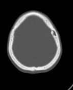
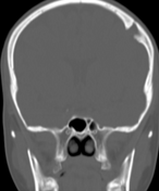
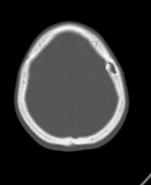
CT of the same patient done few days later revealed:
left parietal bone defect with smooth and sclerotic margins
right maxillary sinusitis with hyperdense metallic foreign body likely migrated dental implant
Case Discussion
This is a case of incidentally seen left parietal calvarial lesion in a patient complaining of headache. The MR appearance, particularly the high signal on DWI, raised the possibility of intradiploic epidermoid. The CT finding of a sharply demarcated calvarial bony defect is also in keeping with epidermoid. Furthermore, CT confirmed odontogenic right maxillary sinusitis which could explain the patient's headache.