
Presentation
Lump on the lateral aspect of the right arm. Bluish tinge to skin.
Patient Data
Age: 9 years
Gender: Female
From the case:
Intramuscular hemangioma
Download
Info
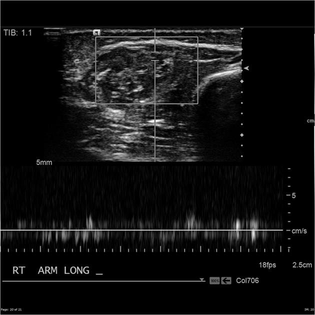
A large mass is situated within the muscles of the lateral aspect of the right distal upper arm. It has numerous tubular internal hypoechoic areas that demonstrate flow. Features consistent with a vascular tumor.
From the case:
Intramuscular hemangioma

Download
Info

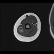
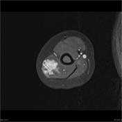
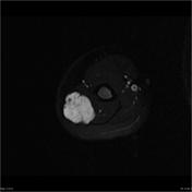
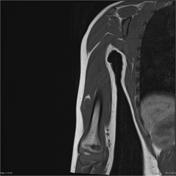
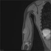
The mass demonstrated on US is again visualized as a high T2, low T1, enhancing lesion. It contains fatty elements (high signal rim on T1 which surpasses on T1 fat sat) and features are consistent with an intramuscular hemangioma.


 Unable to process the form. Check for errors and try again.