
Presentation
Left volar proximal forearm region lump for the last 5 to 7 years. No local pain.
Patient Data
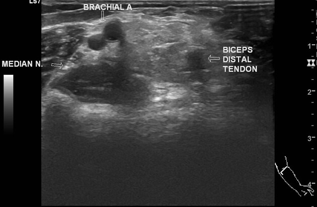
There is a well-defined hyperechoic lesion in the intramuscular/intermuscular plane of the proximal forearm. Internal linear striations are present without calcification/cystic change/vascularity. The lesion showed mild compressibility. The proximal end of the lesion is lies between the brachial artery and biceps distal tendon. The patent ulnar artery is displaced posteromedially by the lesion. The patent radial artery passes anterior to the lesion. The superficial sensory branch of the radial nerve passes the anterolateral side of the lesion.
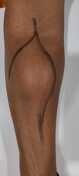
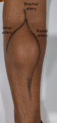
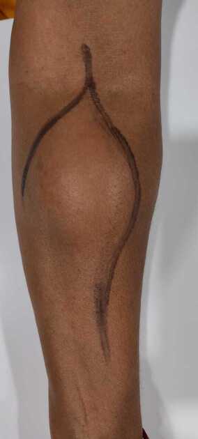
Skin marking of brachial, ulnar, and radial arteries was done to guide a proper surgical incision site avoiding the arterial injury.

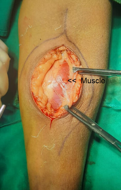
1st photo: shows retracted skin, subcutaneous fat, and muscle covering the lump
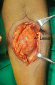
2nd photo: shows the lesion
Image courtesy: Dr Niraj Patel (operating surgeon)
Case Discussion
A female presented with a slow-growing, painless, soft lump lesion in the volar proximal forearm region. The ultrasound features favor lipoma.
Surgical exploration showed an intramuscular lipoma which was excised. Histopathology confirmed the clinical and radiological diagnosis of the lipoma.
Intraoperative photos courtesy: Dr Niraj Patel (operating surgeon)


 Unable to process the form. Check for errors and try again.