Presentation
Left breast and axillary palpable masses on physical exam.
Patient Data
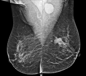
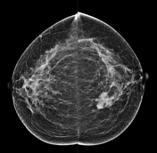


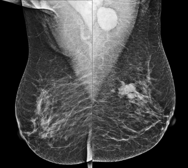
An irregular hyperdense mass with spiculated margins is noted in the medial central part of the left breast, causing surrounding parenchymal distortion (BI-RADS 5). A small satellite lesion is also observed.
An enlarged lymph node is evident in the left axillary region.
The patient underwent an ultrasound-guided core needle biopsy, and the histopathology evaluation confirmed invasive breast carcinoma of no special type. Then, the patient went to have a left partial mastectomy, ipsilateral axillary lymphadenectomy and adjuvant chemoradiotherapy.

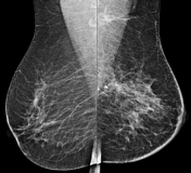
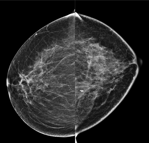
Mild asymmetry is present in the breasts' shape and size due to the left partial mastectomy and ipsilateral axillary lymphadenectomy. Parenchymal distortion with surgical clips is seen in the surgical site without frank signs in favor of local tumoral recurrence. Mild skin thickening is also evident on the left side as post-treatment changes. (BI-RADS 2)
Case Discussion
Invasive breast carcinoma of no special type, previously known as invasive ductal carcinoma, not otherwise specified, is the most common type of breast cancer and often presents as an irregular, hyperdense, spiculated mass with or without calcifications that cause surrounding parenchymal distortion.


 Unable to process the form. Check for errors and try again.