Presentation
Pain in right groin without any history of trauma or positive clinical findings of infection.
Patient Data
![](https://prod-images-static.radiopaedia.org/images/41493254/37f667a1575caa9aede6b64c8af7b6_thumb.jpeg)
![](https://prod-images-static.radiopaedia.org/images/41493245/3bf4bf2d7bea887334486df311a44f_thumb.jpeg)
![](https://prod-images-static.radiopaedia.org/images/41493246/67417c38ed57916af0b47a48f8fbce_thumb.jpeg)
![](https://prod-images-static.radiopaedia.org/images/41493254/37f667a1575caa9aede6b64c8af7b6_big_gallery.jpeg)
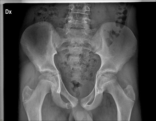
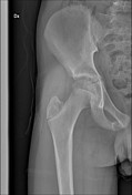
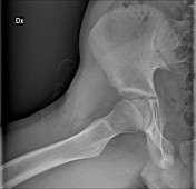
Anteroposterior pelvic radiograph shows enlargement of the right ischiopubic synchondrosis with radiolucency.
![](https://prod-images-static.radiopaedia.org/images/41493283/eed74914ad9f609c3961c8c4086041_thumb.jpeg)
![This study is a stack](/packs/stack-YQKLCKBI.gif)
![](https://prod-images-static.radiopaedia.org/images/41493740/cc4a84bdee6283d7bbe9c0e9dc670c_thumb.jpeg)
![This study is a stack](/packs/stack-YQKLCKBI.gif)
![](https://prod-images-static.radiopaedia.org/images/41493979/322c0a1fc461324ceb1268b13f6bda_thumb.jpeg)
![This study is a stack](/packs/stack-YQKLCKBI.gif)
![](https://prod-images-static.radiopaedia.org/images/41494702/dbf185a54cdc5fa8250a14b06bc5e9_thumb.jpeg)
![This study is a stack](/packs/stack-YQKLCKBI.gif)
![](https://prod-images-static.radiopaedia.org/images/41494441/0aa34a8d14054b17a2fa299b1884d0_thumb.jpeg)
![This study is a stack](/packs/stack-YQKLCKBI.gif)
![](https://prod-images-static.radiopaedia.org/images/41493494/8a7abf8daaa9a750640a78dea5601b_thumb.jpeg)
![This study is a stack](/packs/stack-YQKLCKBI.gif)
![](https://prod-images-static.radiopaedia.org/images/41494464/02f287c1a0f8652f9ee76133be4753_thumb.jpeg)
![This study is a stack](/packs/stack-YQKLCKBI.gif)
![](https://prod-images-static.radiopaedia.org/images/41494211/8cef6162d4099a1f677af8c9caca55_thumb.jpeg)
![This study is a stack](/packs/stack-YQKLCKBI.gif)
![](https://prod-images-static.radiopaedia.org/images/41494823/4dd81381b57a4b89c36f9c25eda05e_thumb.jpeg)
![This study is a stack](/packs/stack-YQKLCKBI.gif)
![](https://prod-images-static.radiopaedia.org/images/41493283/eed74914ad9f609c3961c8c4086041_big_gallery.jpeg)
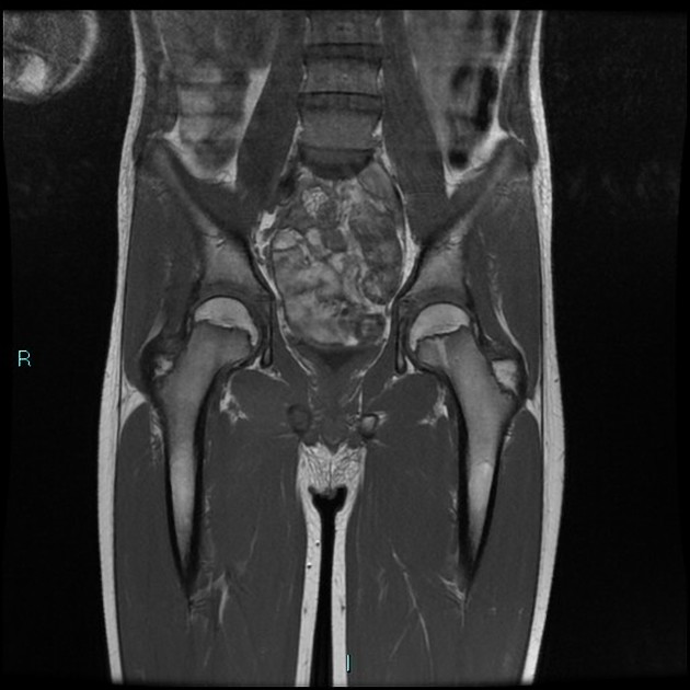
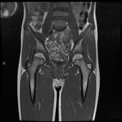
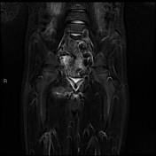
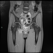
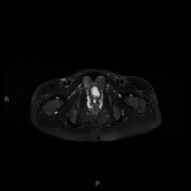
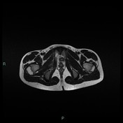
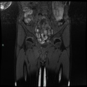
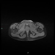
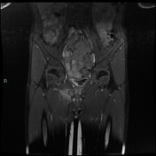
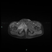
T2 magnetic resonance image demonstrating right ischiopubic syncondrosis enlargement with irregular edges and bone marrow edema; edema of the external obturator muscles and large adductor is associated.
Case Discussion
Van Neck-Odelberg disease is a syndrome characterized by an atypical ossification pattern of the ischiopubic synchondrosis. Its radiological features may mimic stress fracture, neoplasm, osteomyelitis, or posttraumatic osteolysis.