
Presentation
Abdominal pain and distention
Patient Data
Age: 25 years
Gender: Female
From the case:
Krukenberg tumor

Show annotations
Download
Info
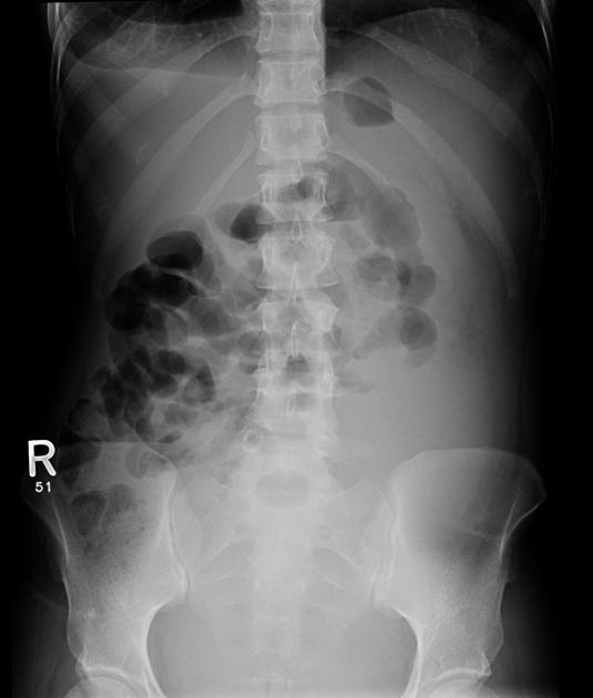
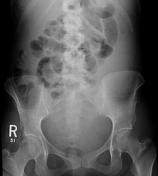
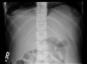
Prominent small bowel loops filled with gas, no air-fluid level, no radiographic signs of intestinal obstruction.
From the case:
Krukenberg tumor
Show annotations
Download
Info
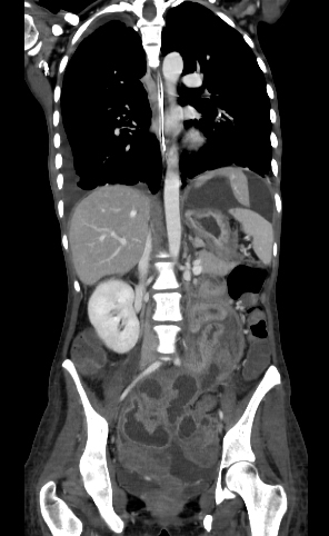
Coronal CT scan demonstrates bilateral ovarian masses which are relatively solid along with moderate volume of ascites
From the case:
Krukenberg tumor


Show annotations
Download
Info
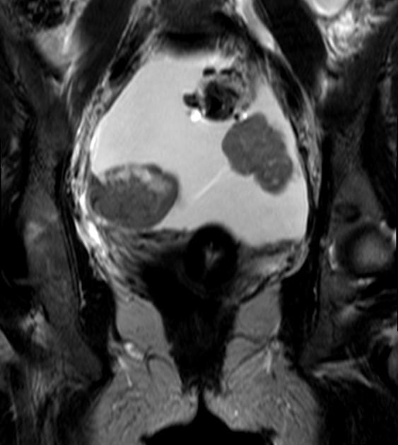
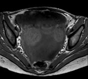
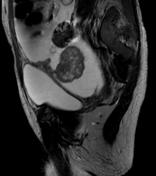
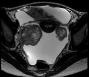
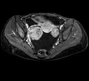
Both ovaries are replaced by lobulated, well defined, heterogeneously high T2, enhancing lesions. Moderate volume ascites see in the pelvis.
Case Discussion
This case illustrates multi-modality imaging of a pathologically proven Krukenberg tumor of gastric origin.


 Unable to process the form. Check for errors and try again.