
Presentation
History of recurrent episodes of jaundice with right upper quadrant pain.
Patient Data


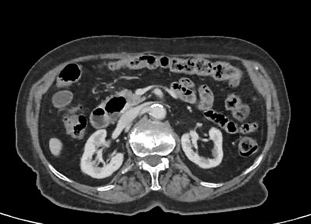
Focal outpouching with a thin wall arising from the medial aspect of the second part of the duodenum (D2), adjacent to the papilla measuring 35 mm, compressing the distal CBD with no wall defect or adjacent fluid or gas collection. The proximal CBD measures 11 mm with mild dilatation of the intrahepatic biliary ducts. Small gallstones are noted, confirmed by ultrasound (not shown).
A simple cortical left renal cyst is noted (5 cm).
Case Discussion
CT features of a duodenal diverticulum arising from the medial aspect of the second part of the duodenum compressing the distal CBD most consistent with Lemmel syndrome.
Lemmel syndrome is defined as obstructive jaundice caused by a periampullary duodenal diverticulum compressing the intrapancreatic common bile duct with resultant bile duct dilatation.


 Unable to process the form. Check for errors and try again.