
Presentation
Left inguinal lump for the last few weeks. Clinical examination revealed a non-reducible lump.
Patient Data
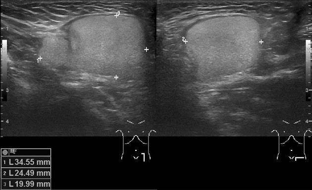
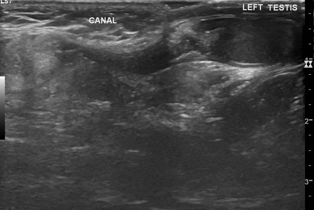
There is a well-defined, homogeneously echogenic lesion in the left inguinal canal; measuring about 40 x 23 x 20 mm. There is no internal calcification/cystic changes/vascularity. The lesion shows lobulation and mild compressibility. There is no inguinal hernia. Both testes are normal in location, size, and echopattern.
Mother noticed recurrence of Lt groin lump about 2 mth after surgery. US was done 4 mth after surgery.
There is a recurrence of the left inguinal mass which is morphologically similar to the preoperative lesion. It is a well-defined, homogeneously echogenic lesion in the left inguinal canal; measuring about 40 x 18 x 18 mm. There is no internal calcification/cystic changes/vascularity. There are lobulations and mild compressibility. There is no inguinal hernia. Both testes were normal in location, size, and echopattern.
Case Discussion
An infant presented with a left inguinal canal lump for the last few weeks. Ultrasound features suggested the inguinal canal lipoma.
Surgical exploration revealed a lipoma in the inguinal canal. Later, histopathology revealed the lesion being a lipoblastoma. It is a rare benign tumor that can recur even after complete excision1.
The mother noticed small bulge at the operative site about 2 months after the surgery and gradually it increased in the size. Follow up ultrasound 4 months after the surgery shows recurrence of the lipoblastoma.


 Unable to process the form. Check for errors and try again.