
Presentation
Right upper quadrant pain and deranged LFTs. Alcoholic, weight loss recently.
Patient Data


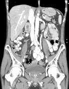
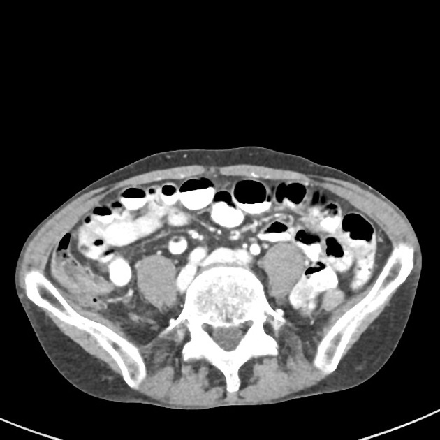
Large non-obstructing mass within the rectum and distal sigmoid colon measuring 7 cm in length, the spine is extensive irregular wall thickening and shouldering in keeping with primary rectal malignancy. Peri-rectal fat stranding is extensive suggesting mesorectal involvement and mesorectal fascia is thickened bilaterally. There is 11 mm upper presacral lymph node and more superiorly a 9 mm lymph node along the superior rectal vessels.
Two ill-defined and possibly contiguous hypodense lesions in the right lobe of the liver, measuring 30 and 20 mm are in keeping with metastatic disease. The segment 8 lesion abuts the anterior branch of the right portal vein which is truncated. The remainder of the portal veins are normal.
Left adrenal gland is bulky measuring 9 x 21 mm which may be metastatic. No other metastatic disease is evident. The lung bases are clear. No bony lesion evident.
Case Discussion
This is a large rectal primary malignancy with mesorectal lymphadenopathy and a right hepatic metastasis with possible intrahepatic portal venous invasion. The classic case for a viva.


 Unable to process the form. Check for errors and try again.