Presentation
Abdominopelvic pain and chronic constipation.
Patient Data
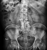
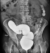
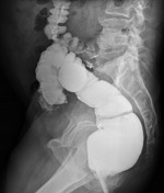
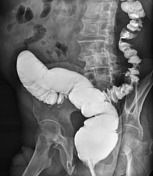
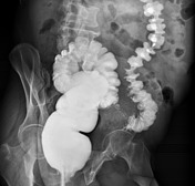
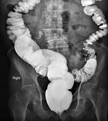
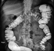
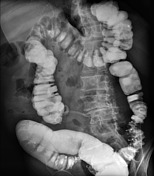
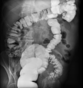
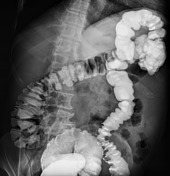
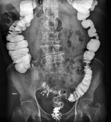
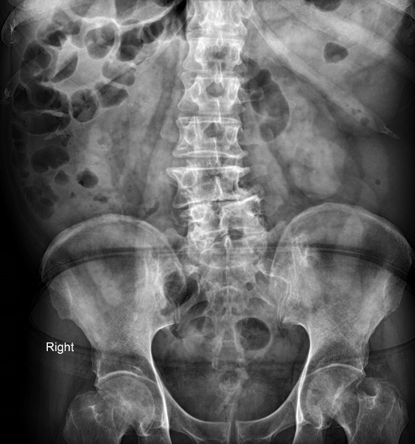
On contrast barium study; dolichocolon is evident. There is short segment stricture with shouldering appearance and apple core sign at sigmoid colon suggestive for tumoral infiltration.
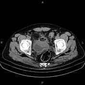

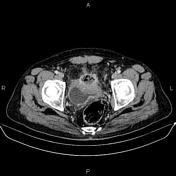

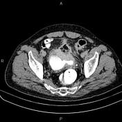

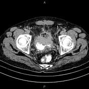


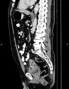

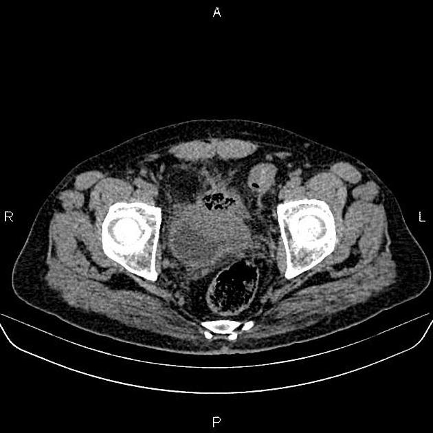
A large mass was seen in anterosuperior aspect of urinary bladder, transgressing its wall and creating perivesical mass. Necrosis with gas bubbles are seen in the mass. In addition,
increased wall thickness with luminal narrowing was seen in the adjacent sigmoid colon.
Four cysts with smooth and thin wall, sharp and distinct marginations, and homogenous water density was seen in left kidney. After IV contrast media, they do not enhance and have no discernible wall thickness.
Additionally, few small stones were seen in both renal calyces.
Case Discussion
Urinary bladder mass (pathology proven carcinoma) with internal necrosis and local spread .


 Unable to process the form. Check for errors and try again.