Presentation
Status post subdural evacuation with positive pressure ventilation became short of breath.
Patient Data

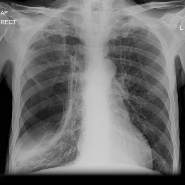
A large lucent area with surrounding compressed lung is noted on the right without convincing shift of the mediastinum.


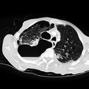
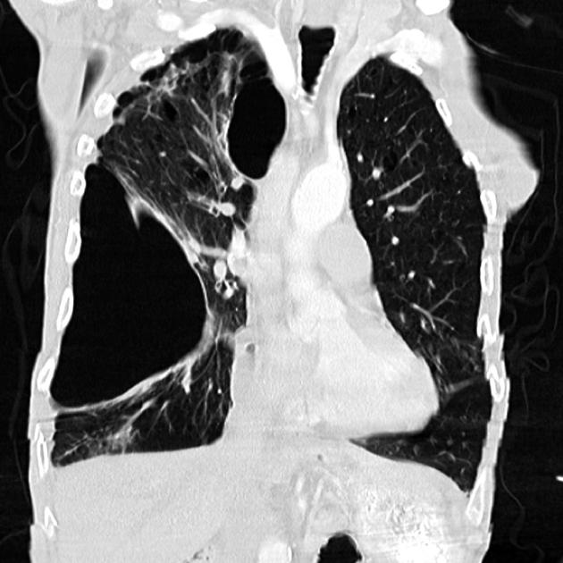
Single coronal and axial CT images demonstrating extensive adhesions at the right apex with a pneumothorax.

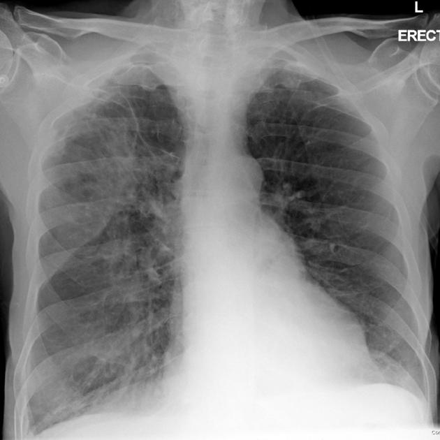
The preoperative chest x-ray demonstrates extensive scarring, particularly in the right apex.
Note: This case has been tagged as "legacy" as it no longer meets image preparation and/or other case publication guidelines.
Case Discussion
Loculated pneumothoraces can be challenging to diagnose.


 Unable to process the form. Check for errors and try again.