
Presentation
Status post subdural evacuation with positive pressure ventilation became short of breath.
Patient Data
Age: Elderly
Gender: Male
From the case:
Loculated pneumothorax
Download
Info
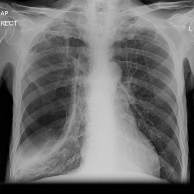
A large lucent area with surrounding compressed lung is noted on the right without convincing shift of the mediastinum.
From the case:
Loculated pneumothorax

Download
Info
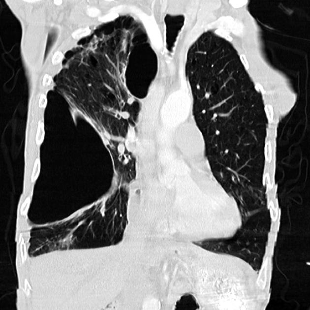
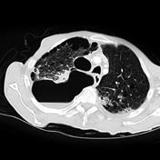
Single coronal and axial CT images demonstrating extensive adhesions at the right apex with a pneumothorax.
Download
Info
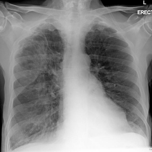
The preoperative chest x-ray demonstrates extensive scarring, particularly in the right apex.
Note: This case has been tagged as "legacy" as it no longer meets image preparation and/or other case publication guidelines.
Case Discussion
Loculated pneumothoraces can be challenging to diagnose.


 Unable to process the form. Check for errors and try again.