
Presentation
Admitted via the emergency department with dysuria and suspected urinary tract infection. A routine chest x-ray was acquired.
Patient Data

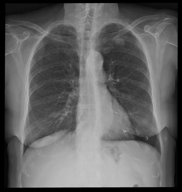
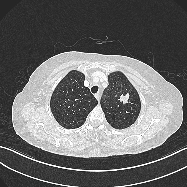
The most striking finding is a round lesion in the left upper lobe measuring approx. 2 x 2 x 2 cm in size.
Besides degenerative changes of the thoracic spine no other pathologies.

Main findings:
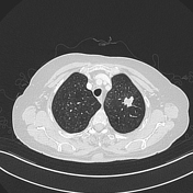
- a highly suspicious mass lesion in the apical upper lobe segment on the left side with a maximum diameter of 2 cm
- suspected satellite lesion in the apicoposterior segment of the upper lobe ipsilaterally. At least two suspicious mediastinal lymph nodes
Suspected bronchus carcinoma results in the following TNM classification: T3, N2, Mx.
Other findings:
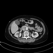
- mild perinephric fat stranding on the right side and to a lesser degree at the left. No ureteral obstruction
- enlarged and inhomogeneously contrasted right ovary
- multiple variable sized cystic hepatic focal lesions
- prominent abdominal aorta atherosclerotic changes
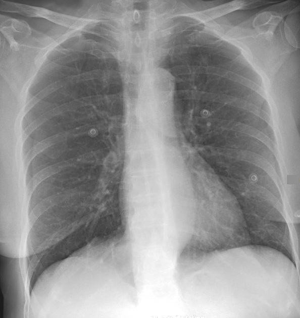
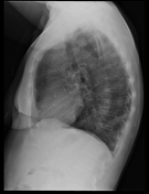
In retrospect, a circular lesion in the left upper field zone is barely distinguishable.
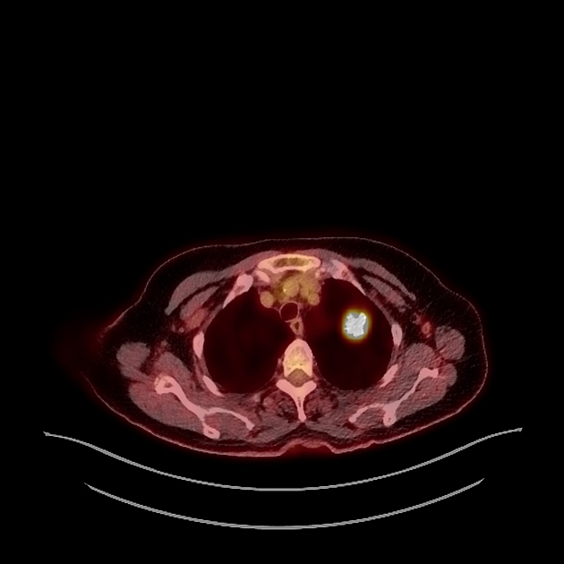
Intense hypermetabolic focus in the left apical upper lobe.
Two hypermetabolic foci in the left upper mediastinum.

Histopathology showed cells of non-small cell lung cancer.
Case Discussion
In retrospect, the newly diagnosed highly suspicious lesion was already distinguishable in the chest x-ray 1.5 years earlier.
Unfortunately, the round lesion in the left upper lobe was overlooked or misinterpreted as of vascular origin. Discovery in this early stage would have resulted in a better patient outcome.


 Unable to process the form. Check for errors and try again.