
Presentation
Neck swelling, back pain with constitutional symptoms.
Patient Data

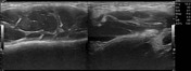
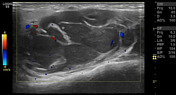
Multi-compartment variable-sized conglomerate hypoechoic lymph nodes are noted, with peripheral vascularity.


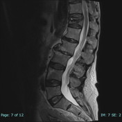
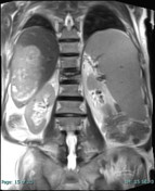
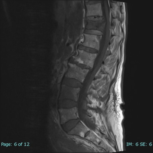
Abnormal bone marrow signal intensity involving most of T12 and L4 vertebral bodies, with extension to the posterior elements at T12 and evidence of hypointense fracture line with decreased vertebral body height, without soft tissue, retropulsion or posterior convexity.
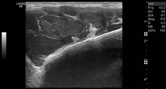
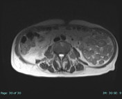
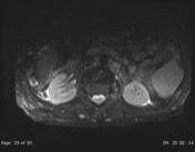
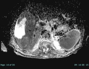
The liver is not cirrhotic, appears enlarged with a large well-defined lobulated lesion involving mainly the right lobe, showing areas of restricted diffusion and central necrosis.
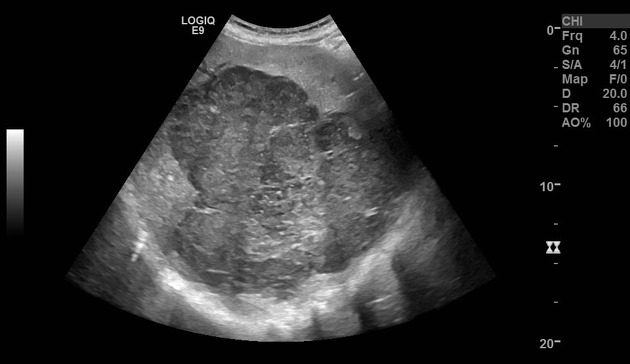
On ultrasound, the liver is enlarged and the right liver lobe shows a heterogeneous lesion with posterior acoustic enhancement.
Case Discussion
The patient presented with neck swelling, back pain and constitutional symptoms.
Neck ultrasound and lumbosacral MRI were requested. The ultrasound showed multi-compartmental suspicious matted lymph nodes.
On MRI, a large hepatic mass was discovered that shows restricted diffusion and necrosis along with hepatosplenomegaly, vertebral pathological fracture and bone marrow infiltration.
In correlation with cervical lymphadenopathy, the infiltrative bony findings are highly suggestive of neoplastic infiltration, with splenic and hepatic involvement, mostly related to lymphoma.
The histopathology report after liver trucut biopsy showed:
Diffuse proliferation of atypical lymphocytes with prominent mitotic activity. The atypical cells are positive for CD20 with a proliferation index of about 80%. CD3 and CD30 are negative.
Diagnosis: Non-Hodgkin's lymphoma, diffuse large B cell.
In conclusion, findings are consistent with non-Hodgkin's lymphoma with hepatic involvement (hepatic lymphoma!), cervical lymphadenopathy, hepatosplenomegaly and bone marrow infiltration.


 Unable to process the form. Check for errors and try again.