
Presentation
The patient presents with acute onset of hypoxia and tachypnea during and post general anesthesia.
Patient Data
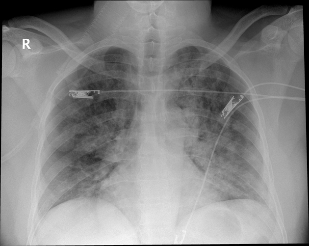
The portable chest X-ray demonstrates extensive, ill-defined, bilateral ground glass opacity and infiltrates. There is a wide differential diagnosis including ARDS, cardiogenic and non-cardiogenic pulmonary edema, and atypical infection including COVID-19 pneumonia and PJP.

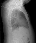
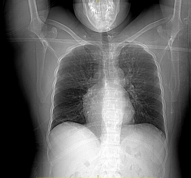
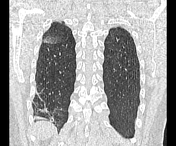
CT scanogram demonstrates persistent infiltrates. There is however significant radiological improvement with supportive fluid, antibiotic and oxygenation therapy within 22 hours of the initial anesthetic.
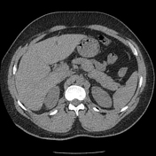

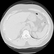

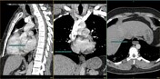
There is a type I sliding hiatal hernia with fluid and gastric contents within the esophagus suggesting gastro-esophageal reflux and alluding to the cause of the acute aspiration pneumonitis experienced during general anesthesia. There is intermittent reflux even between the non-contrast and post-contrast phases in a recumbent position during CT performance as demonstrated within the esophagus between the 2 phases.
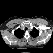
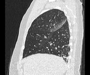
There are typical changes with the posterior and dependent segments of the right upper lobe, right middle lobe, and left upper lobe including the singular segment. These include a combination of tree-in-bud within the left upper lobe, and ground glass opacity within the rest of the affected segments.
There is subsegmental atelectasis within the right lower lobe often a normal post recent anesthetic finding.
There are no aspirated contents within the airways.

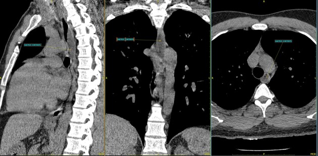
Reformated and annotated images show the type I sliding hiatal hernia and gasto-oespohageal reflux with a variable volume of gastric contents within the upper thoracic esophagus.
Case Discussion
A case of acute aspiration pneumonitis and more specifically aspirated gastric contents (Mendelson syndrome ) during a general anesthetic. The patient underwent a short-duration general anesthetic for the surgical excision of a dermatological mass. The presence of a sliding hiatal hernia and gastro-esophageal reflux during a recumbent position during general anesthesia contributed to gastric content aspiration.
The CT study confirms significant resolution of the pulmonary infiltrates within a relatively short duration of time and obviated the need for aggressive management and intubation.


 Unable to process the form. Check for errors and try again.