
Presentation
Right eye proptosis and decreased visual acuity for one year.
Patient Data
Age: 60 years
Gender: Female
From the case:
Meningioma of the optic nerve sheath

Download
Info

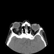
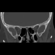
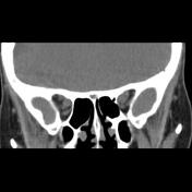
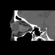
A lobulated margin solid mass lesion in right orbital cavity intraconal encased the optic nerve sheath complex middle to the distal segment containing a few small calcified foci and axial width 20 x 15 mm and height up to 15 mm and with anterior margin in close contact with right eye retrobulbar uveo-scleral layer is seen. Expansion of the right orbital cavity and right side proptosis are seen. A wide sellar cavity with CSF density is noted.
Case Discussion
The case illustrates the non-contrast MDCT features of pathologically-proved optic nerve sheath meningioma. Calcification in this type of meningioma is relatively rare. Empty Sella is also seen.


 Unable to process the form. Check for errors and try again.