
Presentation
Work up for abdominal pain, dysphagia and vomiting.
Patient Data
Age: 60 years
Gender: Male
From the case:
Metastatic esophagogastric adenocarcinoma


Download
Info
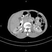
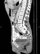
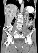
Increased wall thickness due to tumoral infiltration is present at the esophagogastric junction, and gastric cardia and subcardia cause esophageal contrast media stasis.
Several necrotic enlarged lymph nodes are seen at perigastric and upper para aortocaval regions with SAD less than 23 mm.
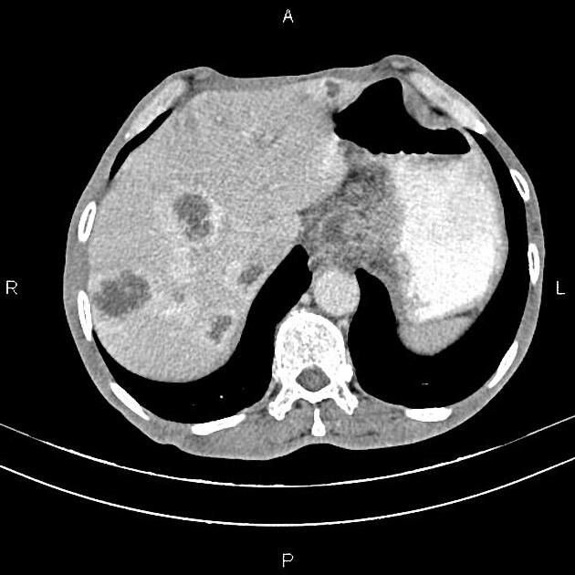
In addition, multiple hetero-enhancing masses are scattered throughout the liver less than 52 mm, inferring metastases.
A few subcentimeter simple cortical cysts are seen in the kidneys.
Case Discussion
Esophagogastric mass; pathology-proven adenocarcinoma with regional and para aortocaval lymphadenopathy and hepatic metastases.


 Unable to process the form. Check for errors and try again.