
Presentation
Metastatic melanoma
Patient Data
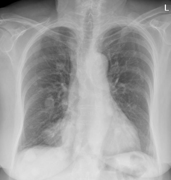
Multiple pulmonary masses/nodules throughout both lungs.
Cardiomediastinal contour is within normal limits allowing for calcified ectatic descending thoracic aorta. No bony abnormality noted. No free subdiaphragmatic gas.


1.8 x 1.2cm well-circumscribed, slightly lobulated hypodensity in segment VIII of the liver. Further subcentimeter hypodensities in segments IVb/V and VI. Right adrenal body hypodense (25HU) nodule. Right hydroureteronephrosis with a transition to normal caliber posterior to the pancreatic head and in the region of dilated right ovarian vein. In the region of the ureteric caliber change, there is ovoid soft tissue density within the ureter. Multiple bilateral subcentimeter renal hypodensities. Pancreas, spleen and left adrenal gland have a normal appearance. Sigmoid diverticular disease. Bowel is otherwise unremarkable.
Hyperdense area of bladder wall thickening (up to 1.3cm) on the right. Right adnexal 5.9cm cystic lesion. No inguinal, pelvic or abdominal lymphadenopathy. Atherosclerotic calcification of the aorta and iliac arteries. No suspicious bony lesions.
Case Discussion
This case demonstrates proven melanoma metastases to:
- lung
- liver
- right ureter (likely)
Incidental findings include:
- left ovarian cyst (possible neoplastic in this age group)
- simple renal cysts (likely)
- sigmoid diverticular disease
- hyperdense bladder wall - may be due to contrast mixing
- right adrenal lesion (metastases vs. adenoma)


 Unable to process the form. Check for errors and try again.