
Presentation
Abdominal pain and progressive distension.
Patient Data
Age: 60 years
Gender: Male
From the case:
Metastatic pancreatic ductal adenocarcinoma


Download
Info
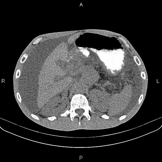
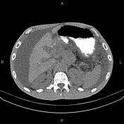
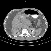
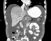
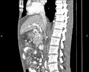
Massive abdominal fluid and omental thickening are present.
A 45× 38 mm ill-defined low-enhancing mass is noted in the pancreatic body and tail that encases splenic vessels and the distal part of the celiac artery. A few enlarged lymph nodes are seen in the vicinity of the diseased segment with SAD less than 12 mm.
Additionally, a few ill-defined low-enhancing masses are seen in the liver less than 25 mm, inferring metastases.
Case Discussion
Pancreatic mass; pathology-proven ductal adenocarcinoma with a vascular encasement, regional lymphadenopathy, hepatic metastasis, ascites and omental thickening.


 Unable to process the form. Check for errors and try again.