
Presentation
Middle aged female with fever, cough, weight loss and chest wall lump.
Patient Data
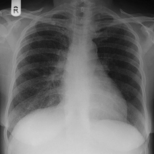
Diffuse miliary nodularity, most pronounced in the right lung.
Small right apical nodule.
Mediastinal contours normal.


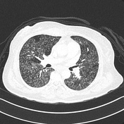

Diffuse miliary nodules throughout both lungs.
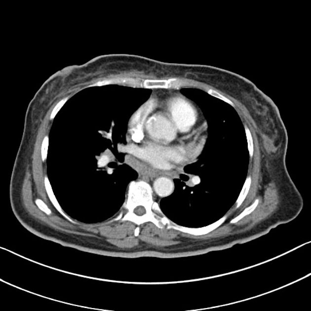
Right anterior chest wall abscess in the parasternal region.
Left paratracheal lymph node.
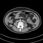
Para-aortic lymphadenopathy in the upper abdomen.
Ovarian dermoid.
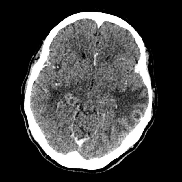
Multiple enhancing lesions throughout the brain with surrounding perilesional edema.
Normal ventricular system and basal cisterns.
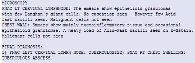
FNAC of the Rt chest wall mass & Lt Cx lymph node
Both the chest wall mass and cervical lymph node contain AFB in keeping with tuberculosis.
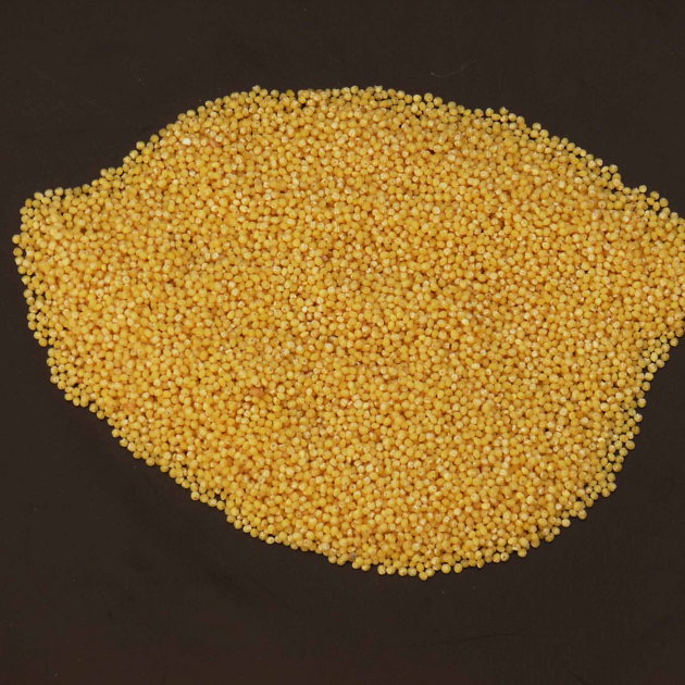
Millet seed from which the aptly name subtype of tuberculosis gets its name. Photo by Ian Bickle.
Case Discussion
There are several causes of miliary nodules, the most clinically profounded being respiratory disseminated tuberculosis - miliary tuberculosis. As with many aspects of good clinical medicine, the correlate with patient's symptomatology is essential.
The appearances of both the chest x-ray and CT chest are classical with innumerable pin-point ( millet seed like ) nodules.
Additional evidence of tuberculosis is evident in the form of the chest wall cold abscess ( the source of the AFFB confirming the diagnosis ) and both supra and infradiaphragmatic lymphadenopathy.
Additional support of multisystem tuberculosis is the widespread intracerebral tuberculomas.


 Unable to process the form. Check for errors and try again.