
Multiple pulmonary arteriovenous malformations with background hereditary hemorrhagic telangiectasia
Diagnosis almost certain
Presentation
Recurrent nose bleeds.
Patient Data
Age: 12 years
Gender: Male
Chest x-ray
Download
Info
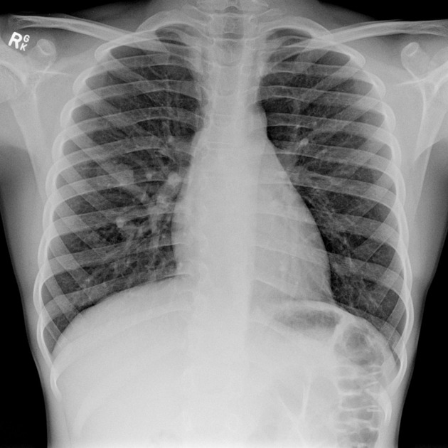
Multiple pulmonary nodules are seen on the chest radiograph, the largest in the right mid-zone. There is a prominent vessel leading to this nodule.

Download
Info

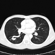
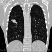
Intravenous contrast enhanced CT demonstrates multiple pulmonary arteriovenous malformations ranging in size from small to moderately large.
Case Discussion
The multiplicity of AVMs and history of epistaxis suggests hereditary hemorrhagic telangiectasia (HHT), also known as Osler-Weber-Rendu syndrome.


 Unable to process the form. Check for errors and try again.