
Presentation
Right hip pain.
Note: This case has been tagged as "legacy" as it no longer meets image preparation and/or other case publication guidelines.
From the case:
Myxoid chondrosarcoma
Download
Info
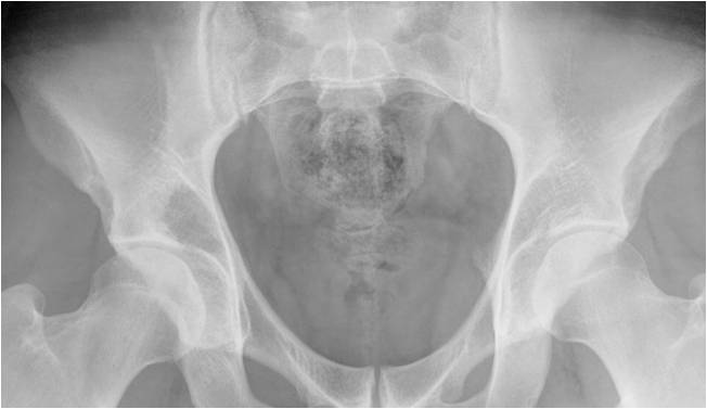
Relatively well-defined area of lucency (with somewhat hazy lateral border) in the upper aspect of the right acetabulum. No obvious calcification within. No septation. The abnormality does not have a sclerotic border. No definite cortical breach.
From the case:
Myxoid chondrosarcoma
Download
Info
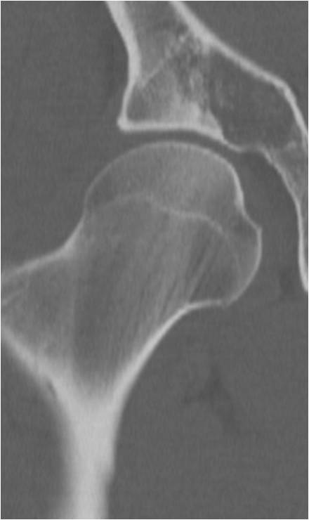
The lesion is better seen on this coronal CT. No definite cortical breach or periosteal reaction. The superolateral aspect of the lesion shows a chondroid pattern of calcification.
From the case:
Myxoid chondrosarcoma
Download
Info

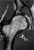
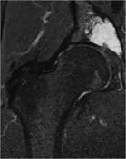
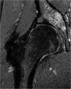
The lesion is of high signal on T2, low on T1 and shows heterogenous contrast enhancement.
Case Discussion
Pathology confirmed myxoid chondrosarcoma.


 Unable to process the form. Check for errors and try again.