
Presentation
Incidental pulmonary nodules discovered on chest radiograph. The patient is asymptomatic.
Patient Data
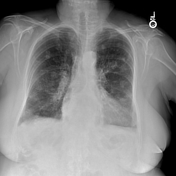

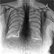

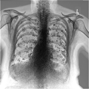
Multiple differently sized pulmonary nodules, some of which are calcified as seen on the dual-energy subtraction radiograph.
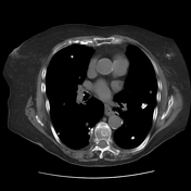

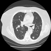

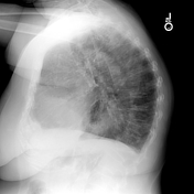
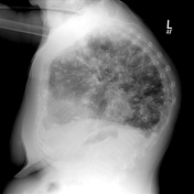
Numerous, irregular pulmonary nodules of varying sizes and varying degrees of calficiation. Some of the nodules are cavitary. They are distributed randomly throughout both lungs.

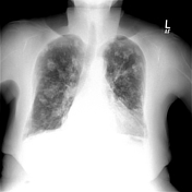

Numerous calcified pulmonary nodules are increased from 7 years prior. Interval radiographs (not shown) show slow progression in size, number, and degree of calcification of the nodules.
Case Discussion
This is a pathologically proven case. Biopsy of one of the lung nodules revealed amyloidosis. The slow progression over seven years also is consistent with the diagnosis.
Differential diagnoses of calcifying metastases, granulomatous disease, or inhalational lung disease are not consistent with the findings and slow progression.


 Unable to process the form. Check for errors and try again.