
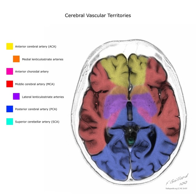
The posterior cerebral arteries are the terminal branches of the basilar artery and supply the occipital lobes and posteromedial temporal lobes.
On this page:
Summary
origin: terminal branches of the basilar artery
course: from basilar towards occiput
-
main branches
posterior communicating artery (not really a branch, see embryology below)
supply: occipital lobes and posteromedial temporal lobes
Gross anatomy
The posterior cerebral artery is divided into four (or sometimes five) segments 8,11. It is worth noting that the definition of the distal segments (P3 and P4) differs between Terminologia Anatomica and routine clinical neuroradiological/neurosurgical articles. This article will only cover the latter.
-
P1: pre-communicating segment
originates at the termination of the basilar artery
terminates to the posterior communicating artery (PCOM), within the interpeduncular cistern
origin of the artery of Percheron variant (supplying the bilateral medial thalami)
-
P2: post-communicating segment
-
from the PCOM around the midbrain
P2A (anterior): sub-segment courses through the crural cistern
P2P (posterior or ambient): sub-segment courses through the ambient cistern
terminates at the origin of the lateral occipital artery/lateral temporal trunk, as it enters the quadrigeminal cistern
-
-
P3: quadrigeminal segment
courses posteromedially through the quadrigeminal cistern
terminates as it enters the sulci of the occipital lobe
-
P4: cortical segment
within the sulci of the occipital lobe
e.g. calcarine artery, within the calcarine fissure
-
P5: terminal branches
terminal branches of the calcarine artery and parieto-occipital artery 11
Branches
Location and presence of branches are variable11.
P1
posterior communicating artery (not really a branch, see embryology below)
P2
-
lateral occipital artery/lateral temporal trunk (most reliably present)
anterior temporal branches
intermediate temporal branches (variable)
posterior temporal branches
P3
temporo-occipital artery (also commonly arises from P2)
splenial artery (variable origin as it may also commonly arise from a medial occipital artery/P4 segment)
P4
P5
Terminal branches of the calcarine and parieto-occipital arteries.
Supply
The posterior cerebral artery curls around the cerebral peduncle to provide supply to the tegmentum and majority of the midbrain (excluding the crus cerebri). Branches of PCOM, P1 and P2 supply the thalamus. The artery then passes above the tentorium to supply the posteromedial surface of the temporal lobe and the occipital lobe, and the splenium of the corpus callosum. The visual cortex responsible for the contralateral field of vision lies in its territory. The macular part of the visual cortex often receives a dual blood supply from the PCA and the MCA, which explains the "macular sparing" phenomenon in some patients following a PCA infarct.
Embryology
The fetal posterior cerebral artery arises as the posterior division of the internal carotid artery. These then fuse in the midline to form the superior most of the basilar artery 9. The proximal portion of the fetal posterior cerebral artery then reduces in caliber remaining as the posterior communicating artery. As such, from an embryologic point of view, the posterior communicating artery is a branch of the internal carotid artery even though in a minority of individuals normal flow in the posterior communicating artery is from posterior to anterior 10.
Variant anatomy
fetal posterior cerebral artery: unilateral incidence 13-15%, bilateral incidence 0.5% 9
fenestration: rare
duplicated: rare, fetal origin and normal origin on the same side 6


 Unable to process the form. Check for errors and try again.