
Presentation
This patient underwent upper GI endoscopy and developed an iatrogenic distal esophageal perforation. Since he had cirrhosis, he was not amenable to surgical repair under general anesthesia. As a result, endoscopic stent was placed. Post stent placement, the patient was referred for a swallow study to assess stent placement.
Patient Data
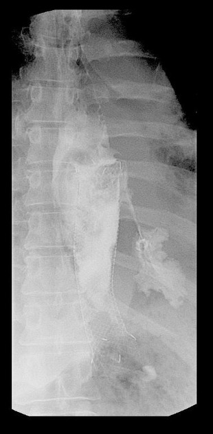
Scout image identifies the radiodense stent in the distal half of esophagus.
First swallow of the water soluble contrast was captured in dynamic cine fluoroscopy. The screen captures reveal extravasation of the contrast from a site immediately proximal to the stent placement likely into the left pleural cavity. The contrast was seen to settle along the contour of the dome of left hemidiaphragm confirming persistent leak.


 Unable to process the form. Check for errors and try again.